Would you be surprised to know that short dogs are more aggressive? Or taller dogs are more affectionate? It seems heavier dogs are more inquisitive and lighter dogs are more fearful too. Whilst we’re not about to take such broad statements at face value, studies have shown that the size and shape of a dog can impact their behavior.
Findings Here
This makes us want to more about the anatomy of our four-legged friends. For that reason, we’ve put together a handy guide in the hope it gives us an even broader understanding of who we share our lives with.
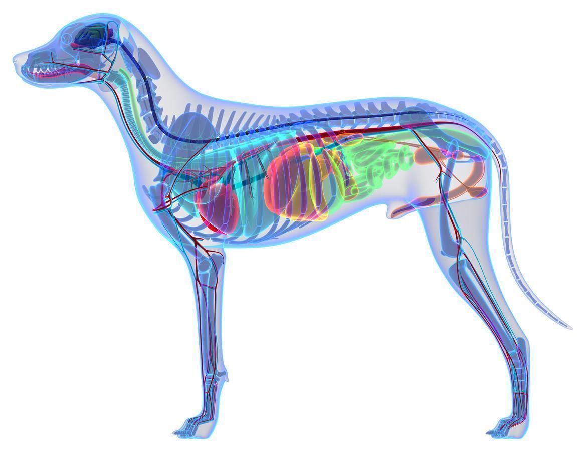
Anatomy of The Dog
With the range of breeds, despite their differences, dogs generally have the same physical anatomy and characteristics which includes their musculoskeletal system. Did you know that dogs don’t have a collar bone, unlike humans; providing a larger stride for running?
Despite their similarities, toy breeds have a skeleton that will mature in around 6 months. Whereas giant breeds can take between 18 months and 2 years to reach full maturity.
Speaking of skeletons, a dog has 320 bones in their body (depending on the length of their tail) and around 700 muscles!
Muscles enable us and our dogs to move. They stabilize our joints and maintain our posture.
Muscle fibers receive electrical impulses from the brain through the central nervous system which tells it whether to contract or elongate; therefore, creating movement.
There are a number of different types of muscles.
Skeletal muscles are connected directly to bones by tendons (elastic type fibers).
Visceral muscles are found inside organs such as the stomach, intestines, and blood vessels.
Cardiac muscle is found in the heart. This muscle is responsible for pumping blood around the body.
Muscle issues:
You may notice muscle atrophy (loss) in dogs who have an injury or developmental issue. Dogs with hip dysplasia or cruciate damage will often have muscle loss in their affected legs.
Injury or over-exertion can often cause muscle spasms, which appear as a localized twitch. This is caused by an interruption in the normal muscle contraction. They can be involuntary or sometimes caused by touch.
Findings Here
Muscles attach to bones via tendons and depending on the breed of dog, they tend to have different types of muscle fibres. You’ve probably heard about slow and fast twitch muscle fibres before? A Lurcher has more fast twitch (anaerobic) fibres in their legs than the Alaskan Malamute who has more slow twitch (aerobic) fibres.
Findings Here
This is of particular interest when we are considering energy usage and systems.
The currency of energy is adenosine triphosphate (ATP). ATP is found in all cells in all the body, but as it is a large molecule, not so much can be stored. To restore ATP there are three relevant energy systems.
ATP-PC
This is where the body uses all the ATP it has stored in its cells. This is the simplest energy production process; and if we were to consider it in human terms, this is the system that your 100m sprint would utilise (or our speedy dogs with their fast twitch fibres).
Glycolytic System
This system runs on glycogen, which is the storage form of carbohydrates in animals. In human terms, this system provides moderate power and moderate duration. Both the ATP-PC and Glycolytic system are anaerobic, meaning they don’t require oxygen to product ATP.
Oxidative System
This system, as its name suggests does involve the use of oxygen to product ATP. This system cannot produce energy as quickly as the other two, but it can produce it continually and for a longer duration. This system can use stored carbohydrates and fats for fuel. In human terms, this would be the system that the marathon runner would access and the system that fuels those slow twitch muscle fibres.
On the subject of legs, let’s look at them in a little more detail.
Just like humans have arms and legs, dogs have forelegs and hind legs. Two thirds of a dog’s body weight is carried on their front legs. Only one third is carried on their hind legs. However, the muscles on their hind legs are larger and therefore stronger!
The foreleg consists of a shoulder, elbow, ulna, humerus radius and wrist. Many large breeds can suffer with elbow dysplasia, where there is abnormal development in the joint. The most common symptom is lameness. Lesions within the elbow joint often start in puppy hood which is why it’s so important to be mindful of their anatomy and typical gait.
The hind leg can be confusing to some owners, but it has some of the same features as a human. The bone between the hip and knee is the femur. Below the knee is the tibia and fibula. Then we get to the hock. You’ve probably heard this mentioned more in horses. The hock is like the human ankle. As with the elbow, many large breeds suffer with abnormal development in the hip joint, known as hip dysplasia. Again, lesions can start in puppy hood. Hip dysplasia is often hereditary but there are also environmental factors which can influence the development of this chronic condition including: rapid weight gain/growth, inappropriate nutrition, inconsiderate exercise, and movement during development.
Can Nutrition Support Joint Health
Puppy Nutrition 101
Obesity and Musculoskeletal Health in Dogs
Moving past the hind legs, let’s look at the tail.
The tail isn’t just something which wags to show you they’re happy – it serves a much bigger function. They can be long, short, curly, or flat! The tail is an extension of the spine, so any injuries are a cause for concern.
The bones of the tail are called vertebrae just like in the spine and they too have discs to cushion the gap between. The muscles and nerves found in the tail contribute to bowel control and movement which is why if a dog ever traps their tail in a door one of the first things to be mindful of is toileting habits.
Happy Tail Happy Dog right?
Not exactly.
There is a condition ironically called happy tail. This is where the dog continuously wags their tail; hitting it on anything they come into contact with. The wall, the coffee table, anything! This can lead to lesions, which due to the nature of the problem (constant wagging), don’t heal. Bandaging the tail often helps, along with keeping the dog in large open plan areas.
A dog’s tail serves many functions including counter-balancing the weight of the body when turning at speed. It is also a crucial piece of the puzzle when observing body language. For example, a stressed or fearful dog will often tuck their tail in between their legs. They may wag it slightly, but very slowly. An aroused or aggressive dog will often raise their tail.
Moving down the leg; after the hock we get to the paw, which as we know is their foot. Their front and rear paws are very similar, just have different names. From their hock (tarsal) joint, there are metatarsal bones which lead to their toes. In the dog’s front legs, from the carpal (wrist) joint, there are metacarpal bones which lead to their toes. The four oval shaped pads are known as digit pads, whereas the large pad in the middle is known as either the metacarpal or metatarsal pad, depending on whether it’s their front or rear leg.
You will also notice a pad slightly further up their front leg; often sticking out. This is known as the carpal pad.
Paw pads are crucial for cushioning of the bones, providing traction and abrasion resistance. If there is trauma or injury to any one of the pads, it can often result in loss of limb function.
Aiding traction also are the dew claws.
These are what you could call thumbs in human terms. They are found higher up than the other toes and don’t touch the ground when your dog is walking. Some dogs only have dew claws on their front legs, some on all four legs – some don’t have any and some have double dews (like the Great Pyrenees). If you ever watch a dog scrambling up a steep bank, you may notice they spread their toes and use their dew claw for grip!
Speaking of claws, let’s have a look at the anatomy of the canine nail.
Many owners are worried about trimming their dog’s nails at the risk of making them bleed. Even more so if the dog has black nails.
The nail consists of a harder outer shell which is usually either pink/white or black. Inside of this shell is a soft cuticle which is known as the quick. This has a blood supply and a nerve. You will notice the quick more easily in light-coloured nails; it is the pinkish area. When nails are trimmed, if the quick is caught; it will bleed and often cause pain.
Dog Skull Anatomy
The main function of the skull in dogs and humans alike is to protect the brain. The interesting thing about dog’s skulls is that they can be different shapes and sizes. Most breeds are categorized into the following dolichocephalic, mesocephalic and brachycephalic.
Dolichocephalic – long headed such as the Afghan Hound
Mesocephalic – skull shape and size somewhere in the middle like the Golden Retriever
Brachycephalic – broader, wide-skulled dogs like the Pug
Interestingly, one studied demonstrated that head shape and size correlated with specific behaviors. For example, Brachycephalic dogs tend to show more interest in humans and appear to be more defensive. Dolichocephalic breeds are less likely to engage in play, but they are also less easily startled and appear to be more resilient in stressful situations.
Findings Here
Another part of the dog’s head which can come in all shapes and sizes are their ears. Some are floppy, some are tall and pointy, and some are just mud and water magnets! Spaniel owners will be nodding in agreement.
Their ears have a similar anatomy to that of a human. The outer ear (the bit you see covered in skin and fur) includes the pinna and the ear canal. The pinna collects the sound waves, sends them down the ear canal to the ear drum. The ear drum makes up the middle ear along with those three little bones; the hammer, anvil and stirrup. We then have the inner ear which includes the cochlea and the vestibular system (which supports balance).
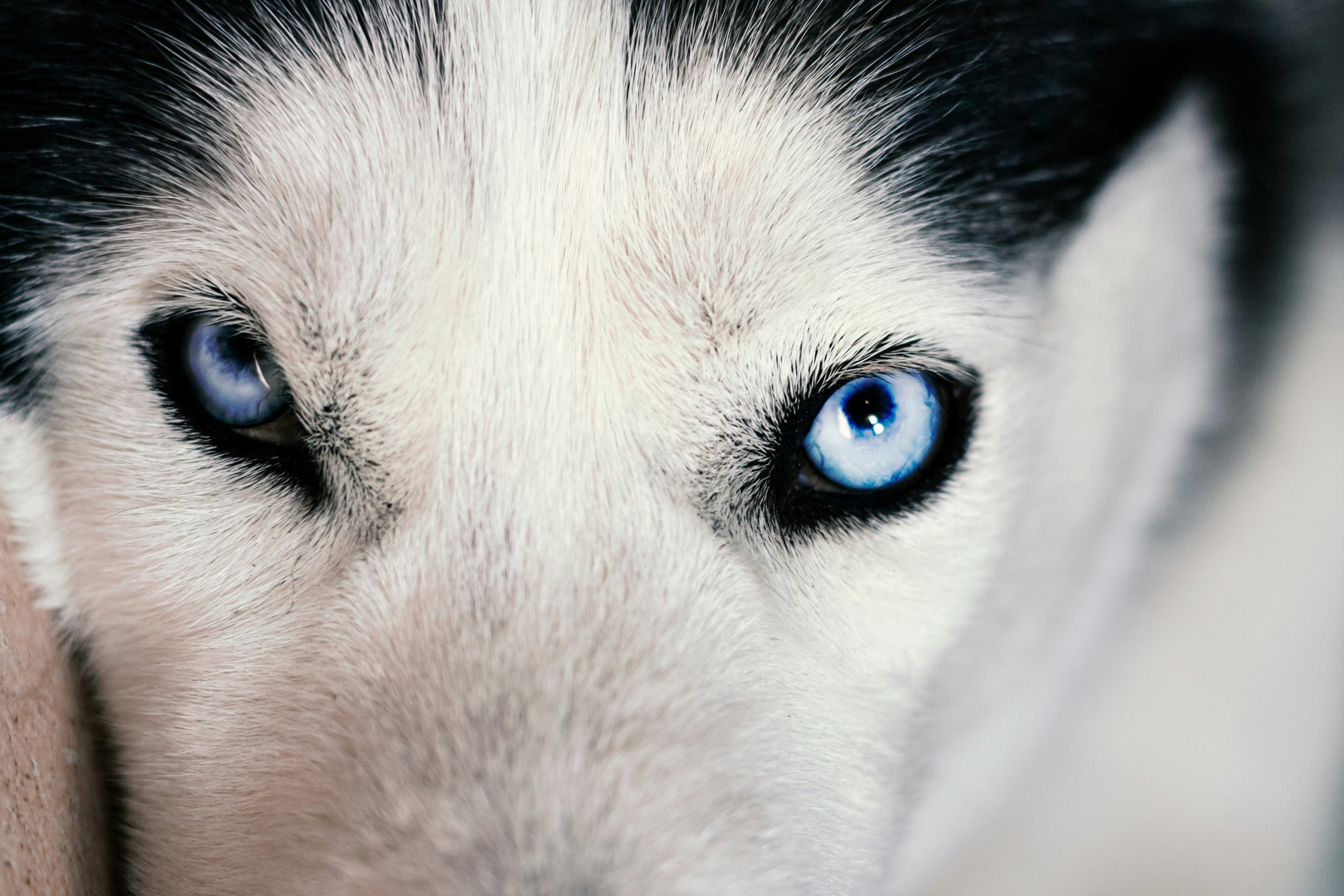
Deafness can occur in dogs for a number of reasons; one of those being pigment related. Deafness is often associated with white-coloured dogs; 30% of Dalmatians are often deaf.
Findings Here
It is common in blue/merle colors in Border Collies and Great Danes too!
Findings Here
Dog Teeth
Also inside of their skull are a dog’s teeth! A grand total of 42 teeth includes 10 molars, 16 pre-molars, 4 canines and 12 incisors when fully grown.
Not surprisingly they have all evolved to serve a particular function.
Canines are designed to grasp and tear.
Incisors are used for cutting or shearing into food.
Pre-molars have a flat biting surface – ideal for crushing.
The molars are the largest teeth, having a flat surface which is ideal for grinding, crushing, and chewing food.
But they don’t have 42 to start off with. Puppies are born toothless; soon growing their 28 puppy teeth. At around 3 months old, these puppy teeth fall out due to the adult teeth pushing through. You may find puppy teeth around your home, but many puppies swallow them.
The tooth consists of a crown, which is usually covered by enamel. Enamel is the hardest substance found in the body. The crown meets the root which is encapsulated by the alveolar bone; known as the tooth sockets found in the jawbone.
Female vs Male Anatomy
Not only are females generally smaller than males in the dog world; they have a few other differences too!
The male has what’s known as a bulbus glandis. This is what’s responsible for the tie that occurs during mating. During copulation, the bulbus glandis engorges with blood which makes it swell. This swelling locks it into the female vagina, demonstrating the typical “knot” or “tie.” When you understand the biology of what happens you can see why trying to separate a male and female during the tie is inadvisable.
As in humans, their testes produce sperm and testosterone. Their prostate produces fluid that helps transport and support sperm. Some of its secretions also have antibacterial properties.
What about the females?
The female reproductive tract includes the ovaries, uterus, vagina, vulva and mammary glands.
Ovaries release eggs and produce reproductive hormones.
The uterus is Y shaped in our dogs. You may have heard it being called the uterine horn due to its shape. When pregnant, the puppies arrange in both horns. At the bottom of the uterus is the cervix, which leads to the vagina. The vulva protects the entrance to the vagina.
The mammary glands are where you will find puppies suckling milk from their Mum! A dog usually has 5 pairs of mammary glands.
Summary
Despite the range of breeds of dogs, they really all have the same basic anatomy. A skeletal system, powerful muscles for movement, ears for listening (or not as the case may be) and teeth for grinding and tearing. Whilst males and females have their obvious reproductive differences, it is still incredible that within a breed they look identical. What is interesting is how different shapes and sizes can be a factor in behavior, especially that brachy breeds tend to show more interest in humans and dolicocephalic breeds are less easily startled. Their muscle distribution also helps us understand their functions and purpose.
The more we know about the pets we share our lives with, the better equipped we are to support their health.
As always if you would like any help in supporting your pet’s health then check out our services below.
Consultations
Thanks for reading,
MPN Team x