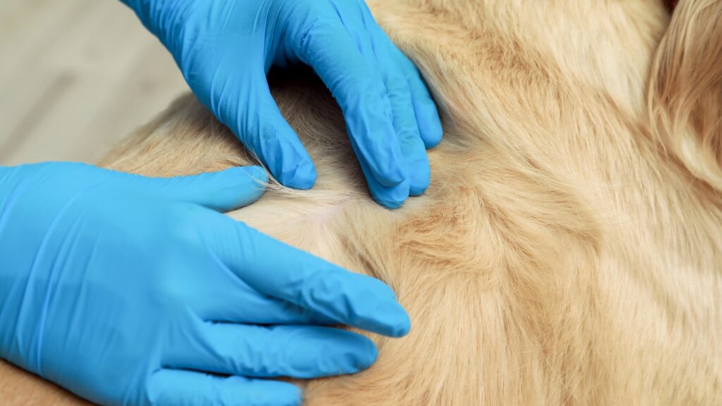
Here at My Pet Nutritionist, we see many dog owners worrying about the condition of their dog’s skin. The appearance of keratosis, and risk of secondary infection can be concerning, but there are things we can do to help those suffering with it. Keep reading to find out what it is, and how we can help those with it. What is Keratosis? Usually referred to as Hyperkeratosis, the condition presents as hardened, thickened, and often cracked and very dry skin. The term ‘keratosis’ stems from overgrowth of keratin in the skin. When too much keratin is produced, the skin becomes less supple. Unfortunately, hyperkeratosis often leads to secondary infection when it is not treated promptly. Those with keratosis will usually find it on the paw pads, and nose. While it can happen elsewhere on the body, these are the most common places it is found in our dogs. Many owners ask if keratosis is painful for their dog. Unfortunately keratosis can be very sore and uncomfortable for them, especially those with cracked skin, and secondary infection. The areas can become very inflamed too, which adds to the pain sensation. Causes of Keratosis There are various factors which can lead to your dog getting keratosis. Let’s have a look at some of these. Age is a huge factor in those with keratosis. It is commonly seen in older dogs, of any breed, but it is most commonly seen in elderly cocker spaniels, boxers, beagles, basset hounds, and various bull breeds. Findings Here Infectious Disease Survivors can be more prone to keratosis, specifically those who have had a systemic infection (an infection which affects the whole body), such as leishmaniasis and distemper. Internal upset in the body, very often presents externally as a skin reaction. The skin is very often an outward sign of inward stress. Findings Here Findings Here Systemic Autoimmune Disease such as systemic lupus and pemphigus foliaceus can also lead to keratosis, again, as an outward sign of inward stress. Findings Here Genetic predisposition is a very common cause of keratosis. It is incredibly important not to overlook lineages/parentage of your dog. This is obviously not easily achievable in most rescue dogs, but if you are planning on buying a puppy from a breeder, a history of skin related issues in the breeding stock is very worth asking for. Labradors are particularly prone to hereditary keratosis of the nose, and usually starts between the age of 6 and 12 months, with recurrent flare ups throughout the dog’s lifespan. A lot of healthy individuals who have no other visible reason for the onset of keratosis, will be diagnosed with having idiopathic (no identifiable cause) keratosis. Findings Here Inadequate Diet Type. The method of feeding being used can also affect the possibility of the dog getting keratosis. As always, we would recommend a fresh food diet, as these are minimally processed, and allow ingredients to provide unaltered nutrition. Zinc Deficiency can cause keratosis. Some breeds of dog, namely the husky and the malamute, cannot absorb zinc efficiently, by genetic predisposition. Many other breeds of dog who grow at a rapid rate, mostly giant breeds, may also struggle to absorb zinc efficiently. One of the main reasons we see in a variety of individuals of many breeds, large and small, is zinc deficiency, leading to keratosis – it is the second most common mineral deficiency, and can be tested for through hair analysis and blood testing at the same time. The lack of zinc can contribute to keratosis as it causes the abnormal production of keratin. Findings Here Findings Here Trauma, particularly to one particular area of the skin, keratosis is common. This is because the cells in the skin, called keratinocytes, react to the repeated trauma by producing excess keratin, with the aim of hardening the skin for protection. This is often why many dogs suffer with keratosis on their paw pads; from repeated walking on hard surfaces. Elderly or overweight dogs who sleep or lay frequently on hard floors often struggle with keratosis due to repeated pressure on certain parts of the body. Findings Here Supporting the Body Externally Recovery from keratosis is a two pronged attack, using internal, and external methods. So what can we do externally to help our dogs suffering with keratosis? As keratosis presents as dry skin, the most important thing to do, is moisturise it. Moisturising the area is imperative to healing keratosis. There are so many natural, and very effective products you can use to moisturise. Coconut oil is one of the most popular, and readily available on the market. Other products include an array of natural snout and paw balms from various companies. Look out for those which do not contain unnatural ingredients – look out for products made from one, or a mix of a couple natural oils such as seed oils and coconut oils, and often a combination of dog friendly essential oils and other seed oils. Apply your chosen moisturising product twice per day, and try to avoid letting your dog lick it off. Supporting the Body Internally The second prong of the two-pronged approach, is internal supplementation, and dietary changes. First we’ll look at diet. A fresh diet would be ideal – these give plenty of moisture, vs dry food which contains very little moisture. Fresh foods are also not ultra-processed, so are much more gut, and therefore skin friendly, as there are huge links with the gut and skin health when we look at the gut-skin axis, for which more information can be discovered in our blog. The vast majority of dry foods on the market have been shown to not meet minimum nutritional guidelines, so deficiencies linked to keratosis are very possible. Using balanced fresh food can be much easier to balance, as we know what is in it. Let’s take a look at supplements. In breeds with a zinc deficiency predisposition, it is important to supplement with zinc. Other breeds shouldn’t require this. The other
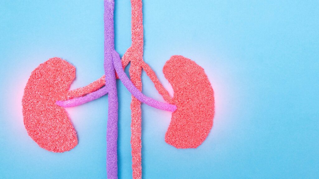
Kidney disease is a common issue we see here at My Pet Nutritionist. In the dog world, there is a lot of confusion over what the diet of a dog with kidney disease should consist of, and if it should be low in phosphorous, or low in protein. We are here to help clear up the confusion! A Bit About Kidney Disease… Chronic Kidney Disease, abbreviated to CKD, happens in 4 stages. In order to understand the need for lower phosphorous and protein, we need to take a little look into the four stages. Stage 1 At this stage, there is no build up of toxins in the bloodstream, unlike in later stages. With often normal blood and urinalysis results, dogs with stage 1 kidney disease often have no clinical symptoms. This makes it very difficult to diagnose early on. Stage 2 During stage 2, toxins begin to build in the blood, and the kidneys begin to lose their function. When the kidneys begin to decline, they often begin to leak protein into the urine, which would give a higher protein reading in a urinalysis than is expected of a healthy individual. Blood pressure in these individuals may rise, but otherwise, clinical symptoms are rare at this stage. Stage 3 Once the diseased has progressed to stage 3, clinical symptoms are common, and include excessive drinking and urination. Following urinalysis, protein levels will be much higher, and blood pressure will further increase. Stage 4 Due to the degradation of the kidneys at stage 4, both protein and creatinine levels are high. Protein levels in the urine become incredibly high, and the blood pressure would be very high. Conventional treatments Upon diagnosis of CKD, your veterinarian may wish to flush the kidneys – this aims to remove toxins. This can help resume normal kidney function, but may not work for all dogs. A lower protein and phosphorous diet will likely be recommended. This diet should not be acidic either! We don’t want phosphorous to flood the bloodstream , so many vets with recommend a phosphorous binder, which is a drug designed to stop phosphorous from entering the bloodstream. Due to raised blood pressure in those with CKD, blood pressure medication may be prescribed. As a last resort, once all other medical avenues have been explored the vet may decide to perform kidney dialysis. What Type of Food Should We Feed Your vet will likely recommend a ‘prescription’ renal diet. These come in both wet and dry variations. Wet or fresh food contains 65-75% moisture, and dry food only contains 8-10% moisture. With the level of dehydration when feeding dry food, it can impact the kidneys further, and is not something we recommend. If using a veterinary diet, it may be a better option to go for the wet version. With this in mind, a fresh diet, tailored to suit a dog with kidney disease, would be the absolute best option. A balanced recipe, with low phosphorous and low-medium protein, would be ideal. We have two fantastic balanced recipes, ideal for those suffering with kidney disease in stages 1 and 2; one tripe based, and one beef based. For those with later stage kidney disease, a consultation is necessary. Where raw is a fantastic diet in general, we would recommend feeding a cooked diet in the event your dog is diagnosed with kidney disease, as cooking further reduces phosphorous. To learn more about kidney disease, and how other lifestyle changes can help those with CKD, please read our blog here! Why Should We Feed a Low Phosphorous Diet? People often wonder what the link is between phosphorous and the kidneys, and therefore why it should be fed in very low quantities to dogs who have kidney disease. When the kidneys are damaged, they struggle to remove phosphorous from the blood. While phosphorous is an important nutrient for strong bones and teeth, as well as muscular recovery, in excessive amounts, it can affect bone health, and cardiovascular health. Studies show that greater excretion of phosphorous in the urine reduces the risk of cardiovascular disease. When the kidneys are functioning as normal, excretion of phosphorous is very normal; but when the kidneys are not able to function as normal, urinary excretion of phosphorous reduces due to their inability to remove it from the bloodstream, which leads to a greater risk of cardiovascular disease. Findings Here When looking at bone and tooth health, we need to consider the calcium-phosphorous ratio. When these are balanced properly, they work together to form strong bones and teeth, and help keep them in tip-top condition. Unfortunately, when there is an imbalance, and there is too much phosphorous in the body, calcium is pulled from the boned and teeth. Removing calcium from the bones and teeth causes them to become softer and weak. Findings Here Findings Here Why Should We Feed a Low Protein Diet? As previously mentioned, one of the main symptoms of kidney disease, is having high protein levels in urine. Studies show that high protein intake can be linked to proteinuria; protein in the urine. When a dog has low functioning kidneys, consumption of protein in ‘normal’ amounts causes immense stress and pressure on the kidneys, which then adds to the build-up of toxins in the blood. Not only does the amount of protein consumed affect the kidneys, but also the type, and quality of protein. Some of the best options for a protein for those suffering from kidney disease include beef, tripe, chicken, eggs, and fish. It’s very important to feed a diet still containing protein, however. Feeding too little can cause muscle wastage, slower healing from injury, cognitive inabilities, and slower metabolism. Aim to feed a low-moderate amount of protein, as part of a balanced recipe for kidney disease patients. Findings Here Findings Here If your dog has kidney disease, especially in the later stages, we would highly recommend booking in with one of our team for a consultation to help keep your dog
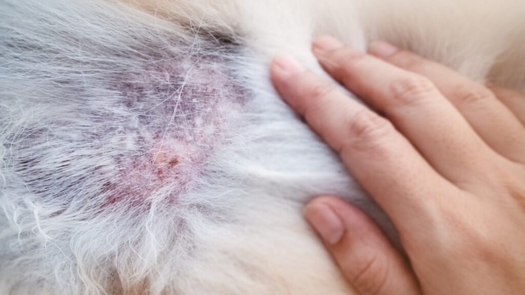
Here at My Pet Nutritionist, we see lots of cases of Hotspots. Those seeing a hotspot for the first time can be worrying; those who deal with them regularly may feel overwhelmed, and frustrated at their regular reappearance. In this blog we will look at what hotspots are, how they happen, their link to gut health, and how we can support the body to heal them efficiently. What Are Hotspots? Hotspots are scientifically known as Pyotraumatic Dermatitis. Another common name for them is ‘Acute Moist Dermatitis’; this name gives more of a visual impression of what you can expect to see of a hotspot case. They present as red raw, moist areas of hairloss, and can happen on both dogs and cats. There is no specific place hotspots can happen on the body, and they tend to show up very quickly. Often confused with ringworm, hotspots are wet in appearance, and often more open/raw looking than ringworm. Ringworm also tends to occur in patches, as opposed to one single spot. Are they sore for your pet? Yes! Hotspots can be very sore for your pet, so it’s important not to leave them. You may notice increased itching and licking of the area, as well as potential behavioural changes due to pain. The sore my ooze liquid, particularly if the hotspot has become infected, which would also bring an unpleasant odour. The fur around the edge of the lesion may be matted. There is no correlation between age, sex or breed when it comes to the likelihood of s dog getting a hotspot. Findings Here What Causes Them? Hotspots are caused by bacterial overgrowth, or are secondary to another underlaying health issue. The biggest trigger for hotspots, is repeated itching of an area on the body. This can make hotspots a secondary health problem to allergies and intolerances, flea infestations and other insect bites, ear infections, grass seeds, stress and anxiety, and poor grooming husbandry when matts are close to the skin. The constant itching and licking of an area makes the perfect growth site for bacteria – moisture and warmth are bacteria’s favourite conditions! Findings Here Findings Here Hotspots and Gut Health As with most skin related problems, there is a huge link with poor gut health. We need to look closely at the Skin-Gut Axis. The skin and gut barrier share many qualities, as they’re both highly vascularised and innervated due to their role in both immune function, and neuro-endocrine function. When looking at the inside of the gut and the surface of the skin, to the naked eye they may seem worlds apart, but both are covered in epithelial cells which both make direct contact with the corresponding environment (the skin contacts particles in the air, things we touch etc, and the inside of the gut touches everything moving through the gut). The biggest similarity between the skin and the gut, is that they both possess their own microbiome. I’m sure you’ve heard us talking about the microbiome on many occasions; but that is because the microbiome is so incredibly important for health and proper functioning of various systems and organs. The microbiome is made up of all microbes the relevant organ uses, and makes contact with, including good (and bad!) bacteria, viruses, fungi, protozoa and other parasites. Gut Guardian There are many studies which prove positive links between probiotic supplementation and skin quality, which can be found in our blog about the gut-skin axis! Metabolites from the gut are shown to link directly to the skin’s ability to fight off ‘nasties’ which would in turn, reduce the risk of hotspots. An important neurotransmitter called Acetylcholine also plays a role in the functioning of the skin’s barrier, and how it deals with overgrowth of bacteria. Supporting the Body for Healing If your dog currently has a hotspot, it’s very important to treat it as soon as possible, as it won’t go away on it’s own. If the hotspot is oozing a yellow or creamy colour, you will need to seek veterinary attention, however it is important to work on the gut following use of antibiotics your vet may prescribe. Step 1: Shave the Fur Gently shave the fur around the hotspot – this enables you to see the full extent of the hotspot, and gives you a clean area to treat it, without the risk of fur getting into the wound. It also allows the hotspot to ‘breathe’, which is imperative in helping it dry up. Step 2: Clean the Area Using either boiled and cooled salt water, colloidal silver, Leucillin/Dew, or probiotic wound spray, gently clean the area to ensure there’s no bad bacteria on or around the hotspot. Pat it dry gently with a cotton pad. Step 3: Apply Green Clay With a clean, soft brush, such as a clean makeup brush, lightly dust some green clay on the hotspot – green clay is an excellent ‘staple’ for the dog cupboard (come on, everyone has a dog cupboard or drawer)! Green clay helps remove toxin from the skin, and helps dry the wound out. When purchasing your clay, ensure the product is 100% green clay, as some products contain added ingredients, including fragrances, which we definitely don’t want to use on our dogs! If you spot the hotspot getting wet through the clay, pop a little more on. Reapply the green clay once or twice per day, and keep the area dry at all times! Step 4: Prevent the Dog from Licking or Itching It’s important to not let the dog lick the wound, or get it wet. Itching the wound can aggravate it too, so don’t let them do this! You may need to resort to the cone of shame (Elizabethan collar, lampshade, or whatever you might call it!) for a few days to achieve this. Depending on where on the body the hotspot is, you may be able to use a clean t-shirt or sock to help cover it. Step 5: Wait! Hopefully
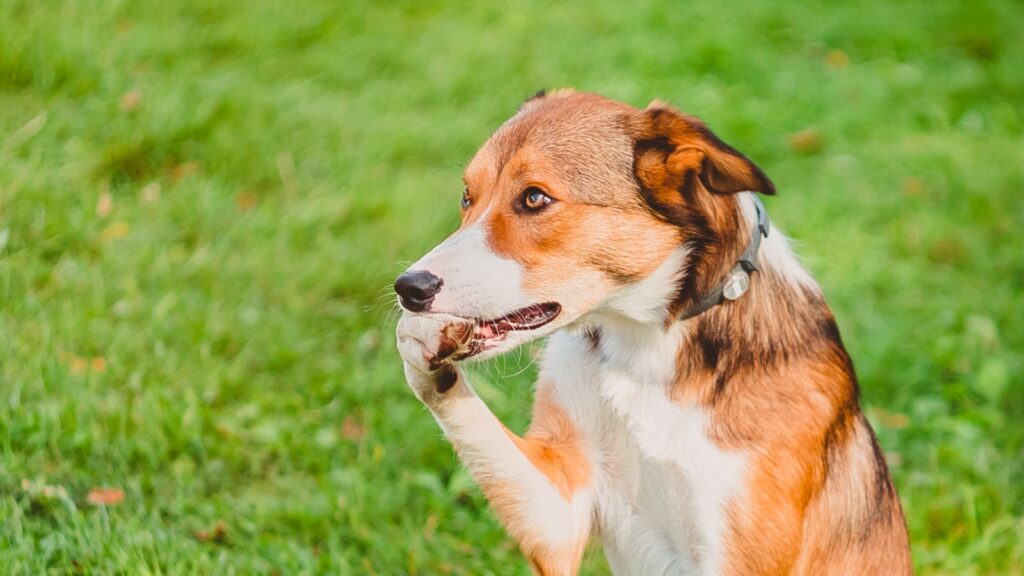
Paw biting is probably one of the most common symptoms we see among our clients at My Pet Nutritionist. It’s daunting when your dog just wont stop! The noise of paw chewing, every owner of a paw biter knows… it can be draining! Our handy guide may help you understand why your dog is biting his paws, and what you could do to help alleviate it. Exposure to chemicals Exposure to chemicals in various aspects of life, can have a massive effect on your dog’s health – especially gut health, which can lead to symptoms such as itchy paws and undercarriage. Let’s discuss the effects flea and worm treatments, household cleaners, and environmental products can have on itchy paws! Flea and Worm Treatments Flea and worm treatments may seem rather an odd cause of paw biting, as flea and worm treatments are administered either orally, or on the skin. These products are so very damaging to the body, both internally, and externally. Not only can they cause chemical burns, and sore skin, as well as neurological problems, they can also damage the gut. A damaged gut can cause a histamine response in the body, which brings with it, symptoms such as excessive itching and licking, including paw biting. We would recommend switching to natural alternatives for flea and worm prevention. Household Cleaning Products It’s important to be mindful of the ingredients used in household cleaning and laundry products. An enormous amount of the ingredients in many laundry products and cleaning products are skin irritants. Try to use as natural products as possible – some natural pet companies stock natural laundry and cleaning products, probiotic laundry and cleaning products, or you can make your own! But my cleaning product states ‘pet friendly’?! Unfortunately, this simply means that it wont kill your pet. Great, but skin irritations are still very prevalent! Dogs make nearly constant contact between their feet, and the floor or soft furnishings, meaning they are more at risk of contact allergies on the feet, causing biting. Environmental Products Navigating around environmental toxins can be extremely tricky. The vast majority of councils in the UK tend to spray weed killer on the streets. These are just one of the environmental products which can contribute to paw biting. Others include patio cleaners, professional astroturf cleaners, grit/antifreeze, and many others. Washing paws after walks is a great idea to help reduce the risk of these toxins affecting the comfort of the dog’s paws. Yeast Yeast tends to have a very familiar symptom, along with the paw biting; a rusty colour on and around the toes. Yeast has two pathways – from the gut, and purely on the skin. Most yeast we see is down to an unhealthy gut, which allows for the overgrowth of candida, however, less commonly dogs may get Malassezia; which is a yeast infection on the skin. Candida Candida is a fungus which occurs naturally on the skin, and in the gut. When it is present in the correct amount, it’s a healthy part of a well-functioning microbiome. The issue comes when candida out competes good bacteria – this leads to a yeast infection. This predominantly occurs in the small intestine, and is aptly named Small Intestine Fungal Infection (SIFO). Overgrowths of candida can be down to exposure to the aforementioned toxins, poor gut health (and therefore a weakened immune system), allergies and intolerances, and antibiotics. As antibiotics not only attack bad bacteria, they also attack the good bacteria, which allows for the growth of fungi such as candida. As yeast thrives on sugar, its important to cut out any high carb, starchy, and sugary foods. Dry food can contain anywhere between 30 and 70% carbohydrates! This means that feeding a balanced fresh food would be the best option. It is also very important to keep on top of, or get to the bottom of allergies and intolerances; running an elimination diet, and healing the gut with mucilage herbs and probiotics is usually the winning combo when getting to the bottom of intolerances. Vaccines, some types of fish such as tuna, and even tap water are contaminated with heavy metals – yeast also thrives on heavy metal exposure, so it’s also important to not use such products, and use filtered water. Working on gut health is very important – you may find our Gut Guardian supplement useful! Findings Here Findings Here Malassezia Folliculitis Affecting only the skin, Malassezia Folliculitis is the skin-specific species of yeast fungus. Often called Fungal Acne, Malassezia causes acne-like pimples on the skin due to the yeast infecting hair follicles. Malassezia can be harder to treat than candida, but thankfully, rarely affects the paws. Findings Here Findings Here You can fund out more about yeast in our blog here! Allergies and Intolerances Unbeknownst to many, allergies and intolerances are very different from one another, and intolerances are often mistaken for allergies. Intolerances are the digestive inability to break down certain foods, specifically proteins. The inability to digest proteins properly, causes intolerances to develop. They can be worked on and improved through gut-work using mucilage herbs, and probiotics such as our Gut Guardian supplement. Allergies are much more severe, and are down to immune modulation – true allergies cannot really be improved. In some cases, allergies are life threatening. An interesting fact, is that in food reactivity cases, on average 10% are true allergies, whereas 90% are intolerances. Food Food allergies and intolerances can affect paw biting, due to poor gut integrity which causes a histamine response by the body. When the body reacts to a sudden influx of histamine, one of the most common symptoms is itching. Irritation can, and often does happen all over the body. Red sores, constant scratching, restlessness, and frustrated panting can all show your dog is itchy. Paw biting is one of these common symptoms too – this shows the dog’s paws are itching, as part of the immune response to foreign particles in the bloodstream. Contact Contact allergies
Here at My Pet Nutritionist, we see a lot of worried puppy parents, struggling with pyoderma. Skin conditions are a very common topic at MPN HQ, so let’s take a dive into pyoderma, what it is, what causes it, and how to support the body with it. What is Pyoderma? The word pyoderma, literally translates to ‘pus in the skin’ (‘pyo’ = pus, ‘derma’ = skin). The condition presents as acne-like spots on the skin, often in the chin area, and around the lips. Pyoderma is a bacterial skin infection, the most common bacteria affecting it being Staphyloccocus intermedius. When there is too much of a specific bacteria present on the skin, the risk of pyoderma is significant. Pyoderma can happen at any age, but is particularly common in puppies – known as puppy pyoderma. Pyoderma can spread between humans and dogs, so it’s important to clean your hands thoroughly when you touch the affected areas on your dog. Findings Here Findings Here Causes of Pyoderma A common contributing factor of pyoderma is environment. Dogs living in warm, humid environments may be at a higher risk than those living in cool, dry environments. Humidity and warmth are essential for bacterial growth. Items in the environment can also contribute to the onset of pyoderma; the biggest culprit being dog bowls! Plastic bowls are particularly good bacterial breeding grounds because they scratch very easily (even if you cant see the scratches to the naked eye), which leaves crevices which are difficult to fully clean, allowing for bacterial growth. When the dog touches the bowl, the bacteria rubs onto the skin, causing pyoderma. An unhealthy gut is also a huge contributing factor to the overcolonisation of bacteria, which can cause pyoderma. 70-80% of the immune system lays in the gut. When the gut is damaged, through the use of chemicals, pest control pharmaceuticals, poor diet or ingredients, or any other cause of gut damage, this can have a severe effect on the body’s ability to get rid of the over-colonised bad bacteria. This leaves the dog more vulnerable to pyoderma. Secondary infection is also one of the major causes of pyoderma. It can be a secondary infection arising from a poor skin barrier, poor gut flora, intolerences to food, and contact allergies from the environment. As well as these common health complaints, there is also a genetic mutation called the Fillagrin mutation – this is much like eczema in humans. Findings Here Breed predisposition may also be a factor in the onset of pyoderma, however more research in this field is needed. Breeds thought to be at higher risk of pyoderma include: Spaniels (ususally lip fold pyoderma) Pekingese Pugs Boxers Bulldogs Shar Pei Read more about genes and skin health here! Finally, stress could contirbute to a dog getting pyoderma. Just like in us humans, stress-spots are very much possible. This is down to the skin having developed it’s own HPA axis. When the body is under stress, the adrenal glands release, and flood the body with stress hormones. This sudden burst of hormones can cause pyoderma to form on the skin. Read more about the skin’s HPA axis here! How to Prevent Pyoderma Prevention of pyoderma is something that may be overlooked by many pet owners, but it can happen to any dog at any age, so it’s something to be mindful of. Using the Right Bowls In order to reduce the risk of bacteria against the skin on the face, and around the mouth, we need to be mindful of the bowls being used for food and water. Glass (modern Pyrex is great, but avoid vintage Pyrex due to the potential for lead contamination!) bowls are a fantastic option, but beware if your dog is likely to pick it up and drop it. Stainless steel bowls are also a good option. Avoid plastic bowls, and be cautious using ceramic bowls, due to the ease of cracking of the glaze, which could then harbor bacteria. Keeping the Gut Healthy As previously mentioned, 70-80% of the immune system lays in the gut, so keeping the gut healthy is incredibly important. There are many avenues to keeping the gut healthy. Feed a fresh diet where possible – dry food is very drying on the gut, and often contains ingredients that can contribute to Leaky Gut. Fresh food, whether it’s raw or cooked using one of our recipes balanced to FEDIAF, is high in moisture, so is better for the gut. Avoid using worm and flea pharmaceuticals – these act a lot like paint stripper on the gut, and contain damaging ingredients, not to mention the risk of seizures, ataxia, and other nasty side effects. Don’t over-vaccinate! The adjuvants used in vaccinations can be detrimental to gut health due to the content of heavy metals. Use a probiotic, and if needed, a mucilage herb. Our product Gut Gurdian is a great choice, combining three mucilage herbs, calming chamomile, and some good quality soil based probiotics. Keeping the Skin Clean Keeping skin clean and dry is very important – focusing on the chin and mouth area, wrinkles/folds in certain breeds, and armpits and groin areas. You may wish to use a hypochlorous based product, of topical probiotic product to clean with, such as Leucillin or Dew (Hypochlorous based), or Provilan LUCAA+ probiotic products. Ensure areas are thoroughly dried. Keep On Top of Allergies Allergy symptoms usually include itching – constant itching of an area will irritate the skin, and leave it open to bacterial infections, especially in the hair follicles. It’s important to work on finding your dog’s triggers, and eliminating them. Keep the Environment Clean Try to keep the dog’s living environment clean! A clean environment, means less risk of bacteria. Less bacteria in the environment, reduces the risk of transmission to a host. How to Support the Body If your dog has pyoderma already, there are some things you can do to support the body through recovery. Let’s take a look at some
As a cat owner, you may have had the joy of dealing with furballs at some point during your cat ownership! The panic setting in when you here ‘that noise’ coming from your cat, the fear of stepping in one when the lights are off… it’s never the most enjoyable part of cat companionship! Here at My Pet Nutritionist, we thought it would be useful to have a short guide on furballs in cats – learn what they are, why they happen, how to help prevent them, and what to do to support the body when the cat has one! What are Furballs? Furballs, are literally as they are called – balls of fur! These balls of fur are forced up through the oesophagus, and expelled through the mouth. They are usually tubular in shape, but can be unformed balls of matted fur, much resembling felt. The scientific name for ‘furball’ is ‘trichobezoar’. Fur is made from a protein called Keratin, just like our hair, and nails. Keratin is indigestible for all animals. How Do They Happen? When your cat ingests a large amount of fur, there becomes a backlog due to it being indigestible. As the amount of fur in the digestive system builds up, the risk of intestinal blockages becomes higher, so the body tries to expel it from the digestive tract. Poor gut motility may make this more difficult. Why Do They Happen? There are many reasons furballs may occur in your pet. Let’s take a look at some of these. General Grooming of Long Fur Longer haired breeds of cat tend to suffer more with furballs, purely due to more hair being ingested during maintenance grooming. The cats tongue acts like a comb – it is covered in tiny hook-like barbs, which act as the teeth of a comb in order to remove knots, and dead fur. Shorter haired cats also ingest fur during grooming, but this tends to be less problematic than those with longer fur. Maine Coons, Ragdolls, Persians, and other long haired breeds are most commonly seen with recurrent furball issues. Over Grooming Over grooming is one of the main problems we see in cats. This is when the cat obsessively grooms itself. While grooming to keep fur separate (and therefore comfortable) is normal, excessively grooming can cause baldness, and massively increases the chances of furballs. Overgrooming can be caused by anxiety in cats, as well as allergies. Stressed cats will often self-soothe by licking/grooming their fur. Those with allergies will be itchy, so the cat will over groom, in an attempt to calm the itch. Pain is also a possible reason for over grooming – in these cases, the cat will usually lick the area of pain. Imbalanced Microbiome The gut microbiome is an important factor in nearly every health complaint, and furballs are no different! Poor gut health can lead to poor motility in the gut. We need gut motility to be fast in order to push fur through, and to reduce contact between pathogens and the gut wall. Poor gut health, and an imbalanced gut with missing or reduced proportions of some beneficial bacteria can contribute to inflammation in the gut. This presents clinically as Inflammatory Bowel Disease (IBD). When the gut is inflamed, it becomes even more difficult due to poor gut motility, to push fur through the intestines and out through the anus in faeces. How Can We Prevent Them? Groom your cat regularly! The more grooming you do with brushes, the less loose fur there is for them to ingest during self-grooming! Try to groom your cat daily, especially if long haired, and also during spring, when the coat is shedding more. Feed fresh! The moisture content of fresh, or high quality wet foods is essential for so many aspects of feline health. Feeding moist foods is also great for gut motility, as the gut requires moisture to move fur along faster. Offering bone broth to those who are unable to eat a high-moisture food may be beneficial to increase the moisture consumed by your cat. Get those omegas in. Omega 3 is very important to reduce inflammation in the body, aiding gut motility, as well as keeping fur soft. The better condition the coat is in, the less shedding that occurs, and therefore the less furballs! Feed probiotics to help the gut microbiome flourish. Keeping the gut microbiome well populated can help gut motility, and help push the gathered fur through the gut. Ensure your cat’s diet contains plenty of fibre! Wheatgrass is a great source of fibre for cats, and can be found in our balanced recipes for cats! Findings Here Supporting the Body If you find your cat has a furball, there are some things we can do to help support the body. Conventional Treatments When you take your cat to the vet, and furballs are found to be the issue, they will often prescribe a form of laxative paste. These often contain artificial sweeteners, malt extract (high in sugar, and made from grains) and other undesirable ingredients, including non-specific ‘hydrolysed animal protein’, or meat meal (ground and dried abattoir leftovers). High sugar dietary components are not suitable for cats, and laxatives may cause other gastrointestinal problems if used regularly. Take a look at some of the natural options below! Fibre While fibre isn’t generally required by cats in large amounts, a little fibre in the diet can go a very long way! Organic Wheatgrass (commonly sold as Cat Grass) is high in dietary fibre, as well as being packed full of vitamins and minerals! It can be offered fresh, or powdered. A tiny sprinkling of Psyllium Husk may also aid gut motility. Mucilage Herbs An important group of supplements for those suffering from furballs, is mucilage herbs. Deglycyrrhizinated Liquorice (DGL), Marshmallow Root, and Slippery Elm are all fantastic choices. Mucilage herbs can help soothe the oesophagus, and the rest of the digestive tract, which is important to keep your cat comfortable during
Here at My Pet Nutritionist, acid reflux is one of the most common issues we come across in our consultations. Dogs with acid reflux may have it for a variety of reasons, from allergies to BOAS, and many other reasons in between. These dogs are often less tolerable of some generally preferred diets, so require a specific diet and supplement regime, which we will discuss here! A Bit About Acid Reflux Acid reflux is formally known as gastroesophageal reflux disease (GERD). When a dog eats, a mixture is formed in the stomach, comprising of stomach acid, salts and bile; this is called chime. Those with acid reflux suffer from the chime mixture entering the oesophagus. Acid reflux is a symptom of many underlying health conditions, and can be very uncomfortable. While acid reflux itself is not life threatening, it can lead to oesophageal ulcers due to repeated inflammation of the lining of the oesophagus, as well as the potential risk of aspiration pneumonia. Symptoms of Acid Reflux include: Bad breath Regular burping and regurgitation Tenderness of the stomach (bowing is the main sign of this) Constipation or diarrhoea Lack of appetite Lethargy Wheezing and dry coughs Weight loss Acid reflux is often caused by low stomach acid, contrary to what many may assume. Proton pump inhibitors are often prescribed under the assumption that the dog has too much stomach acid, however it’s often the exact opposite issue. This may seem an odd concept, however it becomes clear that when the stomach is lacking acid, it is unable to fully digest food at a high enough rate, so the food tends to sit in the stomach, which is then regurgitated. For more information on acid reflux, read our blog here! Diet When is comes to feeding a dog with acid reflux, it’s important not to feed processed foods. Processed foods such as kibble, are very inflammatory, which ultimately reduces stomach acid. A reduction in stomach acid can be a major trigger for acid reflux, as there’s less acid to digest foods. Fresh feeding for acid reflux dogs can be raw, or lightly cooked. Many dogs with acid reflux find it difficult to tolerate raw food, as the food sits in the stomach for a long time; when it sits in the stomach in chime, gasses are released, and the chance of reflux increases. Cooked food is often the best option. When feeding a cooked food, it is incredibly important to follow a balanced recipe, such as our recipes! Sticking to the leaner protein options such as the white fish recipe may be the best option, as feeding fattier proteins may cause more issue for dogs who are unable to digest efficiently. Acid reflux is often a symptom of allergies, so it’s important to get to the bottom of the dog’s allergies, and eliminate trigger proteins from the diet. To achieve this, an elimination diet should be carried out. Why not book in with one of our team for guidance through an elimination diet? Ensure there is plenty of fibre in the diet. Insoluble fibre increases gut and stomach motility, which is essential for those suffering from acid reflux. Some great sources of insoluble dietary fibre are: Broccoli Leafy greens Cauliflower Carrots Berries Findings Here Feeding a good variety of proteins (if allergy constraints allow), and a variety of plant matter can help ensure there are no vitamin deficiencies, which could contribute to acid reflux. In the human world, bariatric surgery patients often suffer with GERD, due to the inability to consume sufficient amounts of vitamins and minerals – nutritional deficiencies can impact our dogs too! Zinc deficiencies are common among acid reflux patients as it is thought that zinc helps protect the lining of the stomach. Findings Here Findings Here Working on getting to the bottom of any allergies and intolerances is important in helping relieve your dog of symptoms too! Allergies and intolerances are largely down to gut health, which causes a knock on effect on acid reflux. It’s also very important to choose the correct supplements to give your dog when suffering with acid reflux. Supplements The first, and most important supplement, or group of supplements we will be looking at, is those that aid gut health. Mucilage Herbs Mucilage herbs include Slippery Elm, Marshmallow Root, and Declycyrrhizinated Liquorice Root. These herbs are excellent for healing the gut, as they coat the digestive system. They not only help heal the gut, but they also soothe the oesophageal tract which is great for those suffering with acid reflux. Our probiotic and mucilage herb blend, Gut Guardian, may be perfect for your dog! Probiotics Probiotics are essential for a healthy gut. They help the gut microbiome flourish, which in turn aids digestion of foods, reducing the risk of chime sitting in the gut. Our aforementioned Gut Guardian supplement contains a great variety of clean probiotics, so may be a great choice for your dog! Findings Here Digestive Enzymes Some dogs, especially our acid reflux dogs, often suffer from poor digestion, due to insufficient amounts of the required digestive enzymes. When lacking such enzymes, food sits in the stomach in the chime mixture, which is them repeatedly regurgitated. Giving a good digestive enzyme may be pivotal to your dog’s recovery, or management of acid reflux. The two enzymes as play in this situation are Pepsin and Trypsin. Findings Here Rhodiola This is a herb, less commonly known in general, which contributes to responding to physical and mental stressors. As well as aiding those with diabetes, cancer, and anxiety, Rhodiola is great at helping the body cope with the physical stressors acid reflux brings. Findings Here Theanine Theanine is a non-protien amino acid which occurs naturally in tea. It plays a role in the prevention of acid refluc flare ups by increasing GABA activity, and reduce oesophageal sphincter relaxations. Together these reduce the symptoms of acid reflux. Findings Here If your dog has acid reflux, and you are unsure what to feed
Here at My Pet Nutritionist, we often help pet owners battle with oxalate crystals/stones in their pets. A diagnosis of any urinary stone can seem daunting, but that’s where we can step in to help! This is a handy guide to the ins-and-outs of Calcium Oxalate stones! What are Oxalate Stones? Oxalate stones, formally known as Calcium Oxalate stones, are formed of microscopic crystals of calcium oxalate. They’re the second most common type of urinary stone found in dogs, second to Struvite stones. If left untreated, oxalate crystals are one of the leading causes of kidneys stones. Males tend to get diagnosed with oxalate stones more often than females, and they’re more common in older dogs than they are in younger dogs. Some breeds are predisposed to Oxalate Stones. These include: Miniature schnauzer Yorkshire terrier Lhasa apso Bichon frise Shih Tzu Miniature poodle Chihuahua Jack Russel Findings Here Findings Here Symptoms of Oxalate Stones There are a number of symptoms caused by oxalate stones, including: Difficulty urinating Bloody or cloudy urine Smelly urine Frequent urination Distended abdomen Lack of appetite Changes in behaviour Lethargy Vomiting If your dog has such symptoms, it’s important to seek veterinary assistance. How Do They Form? Studies show that feeding a diet that causes high levels of urine acidity can contribute to the formation of oxalate stones. Research suggests that urine high in calcium, citrates and oxalates (these changes to urine are largely controlled by diet) can increase the risk of the dog having oxalate stones. Breed, and sex predisposition are huge factors in the formation of oxalate stones, alongside the aforementioned dietary factors. Findings Here Findings Here Diagnosis, and Getting Rid of Oxalate Stones When taken to the vet, the dog will have a blood sample taken, and a full blood panel will be run. A urinalysis will also be performed to check for increased levels of acidity, calcium, citrates and oxalates. If bladder stones are not able to be felt through palpation of the bladder, an x-ray may be performed to work out exactly what we are dealing with. Unfortunately, oxalate stones usually require surgical intervention, as they cannot be dissolved once formed. Like with Cysteine stones, smaller crystals may be able to be flushed out using urohydropropulsion, however as these crystals, untreated, can grow rapidly and cause urine infections, most veterinary surgeons would prefer to operate and remove all crystals as a first port of call. Findings Here Supporting the Body, and Preventing Oxalate Stones There are many ways we can support the body to prevent recurring formation of oxalate stones. Let’s discuss these! Probiotics Studies show that probiotics are effective at breaking down oxalates in the digestive system, so it’s wise to give a good, broad spectrum probiotic every day. Avoid high calcium, acidic, and high oxalate foods It’s incredibly important to keep the pH of the urine as alkaline as possible. There are certain foods we recommend to avoid feeding your dog if they have history of oxalate stone development. These include: Spinach (high in oxalates) Leafy Greens (the darker they are, the more oxalates they contain) Beets (high in oxalates) Citrus fruits (high in citrates and oxalates) Legumes (high in lectin, and oxalates) Beans (high in lectin and oxalates) Nuts and seeds (high in oxalates) Berries (high in oxalates) Increase moisture intake Always insure your dog is drinking plenty of water! A high moisture diet is also incredibly important, be it raw, or freshly cooked. You can also ‘float’ the food, by adding extra water to it. Check out our low oxalate recipe here! Limit sodium intake Another reason to avoid dry foods, is that they’re often high in salt. Salt is very dehydrating on the body, so it’s important to limit sodium intake. Findings Here Findings Here Findings Here Has your dog been diagnosed with Oxalate, of any other type of urinary tones? If the answer is YES, don’t hesitate to book a consultation with one of our team! Team MPN x
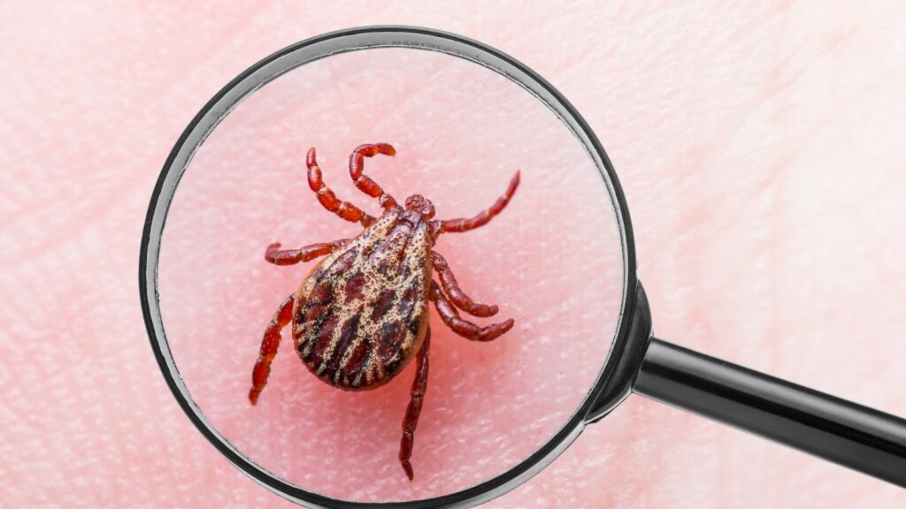
This year, tick infestations seem to be particularly bad. At My Pet Nutritionist, we have had many worried pet owners coming to us, regarding tick prevention, worrying about potential diseases the ticks their dogs are picking up may carry. This guide explains what ticks are, how to identify them, how the severity of tick bites differs depending on your location in the world, what diseases ticks can carry, and how to help prevent tick bites. We will also discuss the dangers of conventional tick treatments, and explain how to safely remove any engorged ticks. (Did anyone get the ode to Fantastic Beasts, and Where to Find Them?! Ticks are definitely not ‘fantastic’ beasts, however!) How to Identify a Tick Ticks are often mistaken for skin tags… or those latched on the stomach area, often mistaken for nipples! Ticks have very small heads, and large, shiny, rounded bodies. They have 8 legs, which protrude from around the head area. Ticks can be a variety of colours, from peachy-nude, to red, to dark brown, grey and black. They can be seen crawling across your pet’s fur, or engorged, in which case, only the large rounded body will be visible, with the legs also visible right next to the skin. We will talk about tick removal later in this article. Ticks in the UK In the UK, our tick population doesn’t tend to carry diseases which are deadly to healthy dogs, unlike other countries which have ticks carrying more severe diseases. We have around 20 species of tick in the UK. The most common species of tick in the UK are Castor Bean ticks (Ixodes Ricinus), Hedgehog ticks (Ixodes hexagonus), and Dog ticks (Ixodes canisuga), though deer ticks are sometimes picked up. Ticks in the Rest of the World Ticks in countries outside of the UK can be much more dangerous than those in the UK, to both humans and dogs. Paralysis ticks are an example – once engorged, Australian Paralysis ticks (Ixodes holocyclus) and the European Red Sheep tick (Haemaphysalis punctata) release a neurotoxin which causes paralysis in the host. Another of the more dangerous ticks is the Lone Star tick (Amblyomma Americanum) which can transmit ehrlichiosis in dogs, which can cause internal bleeding, and death. In humans, the Lone Star tick can cause Alpha-gal syndrome, which is a severe allergy to red meat, and products made from mammals. But What About Lyme Disease? Often carried by Deer Ticks, Lyme disease is the most common potential disease transmitted from ticks. In humans, Lyme Disease is a very debilitating condition, and has a huge affect on one’s quality of life. Humans affected, will likely be on very long courses, or even lifelong medication. Thankfully, Lyme Disease in dogs is much less of a worry! Dogs have a great ability to fight the bacteria causing Lyme Disease. In a study of Beagles exposed to Lyme disease, none of the adult dogs showed any symptoms! Puppies naturally have a less developed immune system, so the puppies in the study had around 4 days of mild symptoms, before their immune system fought off the bacteria! By keeping the immune system strong, we can reduce the risk of symptomatic Lyme Disease in our dogs! Feed fresh, and keep the gut healthy to help keep the immune system strong! Interesting fact: For transmission of Lyme Disease, a tick has to be engorged (attached to your dog, feeding) for 24-36 hours. If removed safely before this period, the risk of transmission is low. Findings Here How To Safely Remove a Tick First we’ll explain how NOT to remove a tick, as this is incredibly important to minimise the risk of disease transmission, and is a common mistake made by pet owners. The cardinal sin in tick removal, is smothering it. Never smother a tick in Vaseline, natural tick prevention, or any other product, and never attempt to burn them off. When you smother a tick, they let go as they are unable to breathe efficiently, however this also causes them to panic. When a tick panics, they regurgitate. Their innards, which could be hosting disease, would be expelled into your dog’s blood stream! If the tick is carrying anything untoward, it would be passed onto your dog! Now onto the SAFE removal of a tick! There are 4 tools you can use! Let’s take a look at each of these! Tick Twisters: These are plastic tools, with a forked end. Simply slide the tick between the forks, right by the skin, so the tick’s head is in the tool, and twist it. The twisting action causes the tick to let go. Tick Keys: These have a large hole, with a very thin opening at the end. Simply hook the tick into the thinnest part of the hole, and pull away from the skin. This pulls the tick from the skin. Tick Lassos: These are pen-like items, with a retractable wire loop at one end. Hook the wire over the tick, and retract it so the tick cannot move. Gently twist, and pull it away from the skin. Tweezers: This is probably the least effective method, but perhaps slightly more accessible if you don’t have a tick tool at home yet (it’s a great idea to add one to your dog first aid kit!). Fine pointed tweezers work best. Pinch the tick with the tweezers as close to the skin as possible, and gently lift the tick away. Be sure to remove the head! Once removed, spray the area with Leucillin, or similar natural antiseptic spray. How to Prevent Ticks There are a number of natural tick prevention methods available. A layered approach is best; an internal product, an external product, and perhaps a repelling collar. Internal products often include herbs such as neem leaf, peppermint leaf, yucca, ginger, fenugreek, lemon balm, and garlic. These all make the dog unattractive to ticks. Fresh garlic is a great internal preventative, in the correct amounts for your size of dog. Do
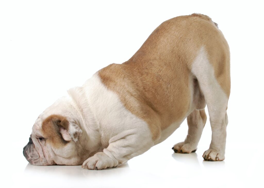
Has your dog had, or been recently diagnosed with gallstones? While they’re not a massively common condition, we do help many dogs with gallstones, here at My Pet Nutritionist. Understandably, owners may be concerned upon news of this diagnosis, so we hope this article helps you understand the condition, and puts your mind more at ease. What Are Gallstones? Gallstones can be found in the gallbladder; a pear shaped organ, located just beneath the liver. The gallbladder is a very important part of the digestive system. It stores bile, which is produced by the liver, and is responsible for digesting fat in the diet. Gallstones are balls of matter, usually made of cholesterol, and hardened bile that form in the gallbladder, blocking it’s ability to secrete bile. Gallstones can be any size from the size of a dust particle, to the size of a ping pong ball! Findings Here Symptoms of Gallstones There are a multitude of symptoms that may present when your dog has gallstones. Let’s take a look at these! ‘downward dog’ yoga pose; this shows a painful abdomen Nausea and vomiting Rapid weight loss Lack of appetite Diarrhoea; can be bloody Jaundice (a yellow tinge to the skin) High body temperature If your dog has any of, or a number of these symptoms, raise this with your veterinarian. How Do They Happen? Poor Digestion Pets with digestive issues may be at risk of gallstone formatsion. Poor digestion can lead to low stomach acid, which can massively affect gallbladder health. In order for the gallbladder to release bile, the stomach needs to be acidic. Lack of stomach acid causes a decrease in bile production. Findings Here Poor Diet Those eating a high fat diet may be at a higher risk of developing gallstones, as the high fat content puts pressure on the gallbladder. When the gallbladder is under the pressure of high levels of fat, it becomes inflamed, and production of the necessary amounts of bile becomes difficult, leading to the formation of gallstones. Feeding a dry food may also contribute to the formation of gallstones, as the fat content of may dry foods, on a dry matter basis, is often high/concentrated. Most kibbles are sprayed with fats and oils at the end of the production line to enhance flavour and palatability. Findings Here Stress Stress hormones have a huge impact on the development of gallstones. When stressed, the liver releases higher amounts of cholesterol, due to the higher production of energy during periods of stress or anger. This increased amount of cholesterol can easily lead to gallstones, as the gallbladder is unable to produce sufficient amounts of bile to digest the excess cholesterol. Findings Here How Are They Diagnosed? If gallstones are suspected in your dog, your veterinarian will want to carry out various tests. An X-Ray will be obtained of your dog’s stomach area, to visually detect any potential masses in the gallbladder, and potential cirrhosis of the liver. A urinalysis will be performed; this is when the vet takes a urine sample and analyses it to look for excess fats in the urine. These results may flag an issue with the gallbladder. Finally, bloodwork will be carried out to look for any abnormal values which may point toward the reduced function of one of the body’s organs. What Negative Effects Can Gallstones Cause? Cholestasis occurs as a result of gallstones blocking the bile duct entirely. This blockage requires surgical removal. When the gallbladder is blocked, absorption of vitamin D and Calcium becomes poor, as well as the aforementioned symptoms. Acute Pancreatitis is also a risk posed to those with gallstones. Gallstones can exit the gallbladder and cause blockages in the bile duct, which ultimately blocks pancreatic enzymes from entering the small intestine. These enzymes are forced back into the pancreas, which creates immense inflammation to the organ, leading to the onset of acute pancreatitis. Findings Here Findings Here What Treatments Are Available? Generally speaking, surgery is usually the main option when treating a dog with gallstones. Some mild cases may get away with having IV Therapy, whereby the dog is hooked up to an Intravenous drip, designed to flush the gallstones from the gallbladder. The patients will usually be prescribed a high protein, low fat diet moving forward… this is where supporting the body naturally comes in! Read on! How Can We Support the Body Naturally? Feed Fresh As with any condition, we always recommend feeding fresh food, here at My Pet Nutritionist; whether that’s raw, or cooked. Balancing the fat content in Fresh food is much than in dry food, and is not sprayed with fats and oils during production, adding to the overall fat content. Fresh food is also high in moisture, and generally better for digestion and gut motility than dry food, making it the ideal option for those with gallstones in their medical history. Feed Low Fat Feeding low fat is also essential for those suffering from gallstones, due to gallstones being solidified lumps of cholesterol. As the gallbladder is unable to produce enough bile to digest the fats consumed in the diet, fatty deposits will begin to build up, which is especially dangerous for the body’s organs. Find our low fat recipes below! Low Fat Fish Low Fat Venison Low Fat Horse Low Fat Kangaroo Dandelion Dandelion has many health benefits for your dog! One benefit being that it’s a fantastic digestive aid. When suffering from gallstones, digestive support is crucial. Dandelion stimulates the production of digestive juices, which are ever so important when it comes to gallbladder health, and gallstone prevention. Dandelion is also a great natural anti-inflammatory. This is beneficial for those suffering from gallstones or poor gallbladder health in general, to reduce the risk of acute pancreatitis occurring. The root of the dandelion plant is also a diuretic, bringing benefits to the liver, which includes increasing circulation in the liver, resulting in an increase in bile production. Findings Here Burdock Root A more unusual vegetable, burdock,
Immunity is no doubt at the forefront of most pet owner’s minds. Everyone wants their pets to live a long and healthy life, so keeping a strong immune system is essential. Here at My Pet Nutritionist, we help many pet owners through their own experiences of disease in their pets, so we thought we would put together this blog on the cornerstones for a strong immune system in your pets. 1. Gut Health Gut health is key to general health of all species, whether it be human, canine, feline, or even smaller furry pets! Virtually all aspects of health can be improved through good gut heath. Around 70% of the immune system is found in the gut! Quite a staggering figure, but a very important fact to keep in mind when it comes to our pets’ health. In an unhealthy gut, the immune system becomes massively impaired, leading to a potential multitude of health issues, including those allergy dogs we so often help here at My Pet Nutritionist. Many things can damage the gut, including chemical flea and worm treatments, poor diets which are processed and/or high in ingredients containing lectin, contact with household and garden chemicals and much more. It’s imperative to gut health, to feed fresh, and limit the exposure to all toxins! Using a mucilage herb for those with damaged guts, helps to heal the gut; these include slippery elm, marshmallow root, and deglycyrrhised liquorice. Teaming these with a probiotic allows the gut to flourish, as a damaged gut will leak the good bacteria which forms a large part of the immune system. Gut Guardian 2. Methylation DNA Methylation is a very important chemical process within the body. This process includes the chemical modification of DNA during replication of cells. Gene expression can be altered through methylation too. So, why is this process an important part of keeping the immune system strong? Various malignant cells, including those for lymphoid cells (produces immune cells B, T and NK) and myeloid cells (related health issues, such as Mast Cells) replicate through a process called hematopoiesis, making these cells ‘hematopoietic stem cells’ (HSCs). Myeloid cells are not ones we want replicating in the body, as they can lead to various types of tumour. Here’s where the process of Methylation comes in to play! Methylation has the ability to regulate HSC differentiation – in other words, it reduces the risk of the Myeloid cells replicating, and forming tumours, but increases differentiation of lymphoid cells. For more information on the types of cells involved in immune responses, read our blog here! Findings Here Findings Here Findings Here 3. Healthy Brain and Endocrine System The Endocrine System is what keeps the body’s organs in good health, through homeostasis. The endocrine system and immune system used to be thought to work independently from one another, but more recently, it was discovered that the two systems work hand in hand to keep the body healthy, and control infection. The immune system uses a mixture of immune receptors and cytokines to fight infection, and the endocrine system uses hormones to regulate metabolism in the body’s organs. As an example, one of the most important regulations aided by both systems, is the metabolism of glucose. Insulin is produced by the endocrine system to regulate glucose levels in the blood, and helps the body to fight infection. Cytokines aid the body during glucose spikes by increasing responsiveness of the peripheral organs to the endocrine system. When the organ becomes more responsive to the signals from the endocrine system, greater amounts of the required hormone can be produced. The brain produces so many important hormones for health of the body, and to help fight a variety of infections, so it is important to keep the brain healthy and free from inflammation. Supplementing your pet’s diet with plenty of omega 3 is a great way to keep inflammation down. To read more about the endocrine system, and the importance of hormones, read our blog here! Findings Here Findings Here 4. Circulation and Lymphatics The lymphatic and circulatory systems are an important part of immunity, and keeping the immune system strong. The lymphatic system is responsible for draining excess fluid from bodily tissues, removing cellular waste, absorption of fat soluble vitamins from dietary sources, and helping to fight infections. The circulatory system works with the lymphatic system to transport the cellular waste and excess fluid around the body, and away from the relative organs. The interstitial fluid (found between tissues) is what provides the body’s cells with important nutrients, and a way of removing any waste from the cells. The lymphatic system acts as a taxi for antigens and antigen-presenting cells to transport them through the body to places of infection. This enables the antigens to fight the infection in question, and expel waste. Findings Here Findings Here 5. Detoxification Finally, detoxification is essential for immune health. The body is exposed to a host of toxins, just by going through daily life. We live, generally, in a very toxic world, so detoxifying the body is essential. Detoxification aids methylation, which as discussed above, is a very important process for a strong immune system. Liver Guard Heavy metals can be picked up and consumed very easily in our pets – traces can be found in food, and heavy metals are also in abundance in vaccinations. Those who have received vaccinations during their lives, whether only one has been given at an appropriate age as a puppy, or they’re given regularly (if given regularly, over vaccination occurs, making the risk of vaccinosis higher; read more here) the likelihood that your pet has heavy metals in the bloodstream is high. Heavy metals disrupt metabolic functions in vital organs, as well as reducing the ability to efficiently absorb vitamins and minerals from dietary sources. Detoxification can be achieved through feeding a fresh, preferably organic diet, minimising exposure to environmental and veterinary toxins, occasionally offering Epsom salt baths, supplementing the diet with milk thistle (this
A diagnosis of Diabetes in your pet can feel quite daunting. We decided to put together this guide on diabetes in pets, to bring all you may need to know about diabetes, and caring for a diabetic pet, into a handy, bitesize guide! What is Diabetes? Diabetes is a condition causing blood sugar levels to become too high. There are three types of diabetes; Type 1, type 2, and Diabetes Insipidus. Type 1 is found in around 0.2 to 1% of dogs, but type 2 diabetes is not found in dogs; it is however, found in cats. Diabetes Type 1, is most commonly found in adult dogs between the ages of 6 and 9. Diabetes Insipidus is another type of diabetes, unrelated to type 1 or 2, but with similar symptoms. Findings Here Symptoms of Diabetes Symptoms of diabetes in pets include: Polydipsia (excessive water consumption; read more here!) Polyuria (excessive urination; read more here!) Increased appetite Weight loss (often rapid) Recurring skin and urinary infections Clouded eyes A dog with diabetes may present with any number of these symptoms, so if your dog is displaying one or more of these, it’s best to have a full vet check. Findings Here What Causes Diabetes? A huge part of the cause of diabetes in dogs, is down to genetic predisposition. Breeds predisposed to the potential to develop diabetes include: Miniature schnauzer Dachshund Beagle Poodle Samoyed Keeshond German Shepherd Golden Retriever Labrador Retriever Bichon Frise Pug Cocker Spaniel Diabetes can also be caused by other ongoing medical problems. It can be a secondary condition to Cushing’s Disease, Pancreatitis and obesity. Those suffering with pancreatitis flare ups are at risk of diabetes due to the damage on the organ, caused by regular periods of intense inflammation. The pancreas secretes insulin which regulates blood sugar levels. When insulin is not able to be produced due to damage to the pancreas, blood sugar spikes occur, which leaves dogs at a high risk of diabetes. 30 to 40% of dogs with diabetes, also have pancreatitis. Obesity, like in humans, is a common predecessor for diabetes as it contributes to insulin resistance. Resistance to insulin, means less control of blood sugar levels. Findings Here Diagnosis of Diabetes To test for Diabetes, your veterinarian will take bloods and urine samples. In the urine sample, they will test levels of glucose and ketones. If there is a significant abnormality in these results, the blood glucose level will be tested. If glucose levels are very high in the blood sample, and found in the urine sample, a diagnosis of diabetes can be made. During testing for diabetes mellitus (Type 1), other significant markers for other conditions may be found, so your veterinarian may start treatment for other conditions as well as diabetes. If your dog’s urine is extremely dilute, diabetes insipidus may be a diagnosis your vet may consider. Findings Here Monitoring, and Medical Intervention Insulin Therapy After diagnosis, the first thing your vet will work out, and prescribe, is the right insulin dose for your dog. The vet will teach you how to give an insulin injection, either by syringe, or VetPen. These will need to be given regularly alongside monitoring, to ensure your dog’s blood sugar levels stay consistent and within a normal range. Findings Here Monitoring Glucose Levels Traditionally, your vet will teach you how to monitor urine glucose levels at home. Urine can be tested using urine strips, just as you would in the human world. The vet may also teach you to test your dog’s blood glucose levels. They will provide you with a portable glucose meter, which you will use in tandem with blood test strips. Bloods may be taken, and analysed regularly by the vet to keep an eye on blood glucose levels and ensure the insulin dosage is correct. More recently, some veterinary practices have been offering the Freestyle Libre device worn by many human diabetics. This is a device inserted into and attached to the skin, which reads blood glucose levels. When the owner needs to check glucose levels, they simply use their accompanying mobile phone application which connects to the device. This is an expensive option, but very fail safe, and easy. Findings Here Findings Here Spaying Females Your veterinarian will recommend you spay your female dog if they are diagnosed with diabetes. One of the female sex hormones, called progesterone, can sadly interfere with insulin usage in the body, which could cause a blood sugar spike. Findings Here Supporting the Body Naturally Diet As with anything, we always recommend feeding a fresh diet. Why is this so important for diabetes sufferers? The carb content in dry foods causes a spike in blood sugar levels. Forcing a raised blood sugar level on a diabetic dog will put a huge amount of pressure on the organs, and can cause hospitalization. When looking at the ingredients list of dry foods, there may be very few ingredients which appear to be carbohydrates; however, these do add up! The carb content of dry foods isn’t specifically listed, only protein, fat, moisture and ash. If you add those up, and deduct from 100, you will get the percentage of carbs in the food. Most dry foods are between 20 and 70% carbs! Three very important components of a diabetic dog’s diet, are fibre, fat, and protein. Fibre, fat and protein help to stabilise blood sugar levels as the consumption of these macronutrients causes the digestion of carbs to slow down, ultimately slowing down their absorption into the blood. Because the absorption of the sugars from carbohydrates is slowed, blood sugar levels stay more controlled, and are more unlikely to spike. Findings Here The moisture level of fresh food, also helps keep the urine suitably dilute, and not concentrated in glucose or ketones. You will need to keep the amount fed each day the same, as this is what your dog’s insulin dose is based upon. If the dog is fed more than normal, the