Rabies is a scary disease, with low survival rates if treatment is delayed. While we don’t see many cases here at My Pet Nutritionist, we do have lots of readers from the USA and other countries with a risk of rabies. It is not a disease present in the UK, unless an imported dog was to enter the country with it. In this blog, we will look at what it is, how it is diagnosed and treated, and how we can support dogs naturally. We will also discuss the rabies vaccine, prevalence of rabies and laws in Europe, Australia and the USA, as well as considerations when travelling from the UK with a pet. Diseases can be viral or bacterial; Rabies is viral, and is zoonotic, meaning it can pass between species, and is not species specific. Rabies is a Lyssavirus; which is one of a group of bullet-shaped RNA viruses causing brain inflammation and rabies-like disease. The most common Lyssaviruses are RabV (Rabies Virus) and ABLV (Australian Bat Lyssavirus), which both have similar symptoms, and both attack the central nervous system. Different diseases are contracted through different pathways, whether it’s an airborne disease, transferred through a cut or bite, or through saliva. In the case of rabies, it is transmitted through the saliva of rabid animals into open wounds like bites. The disease can be fast progressing or extremely slow progressing over a number of weeks, but as soon as clinical symptoms show, the chance of recovery is reduced. Findings Here Symptoms There are a number of symptoms, which change as the disease progresses. In the early stages of Rabies infection, you may notice your dog is weaker in the legs, more lethargic, and has a raised temperature. Once the disease is advanced, symptoms become severe, and affected individuals clinically present with aggression and agitation, complete disorientation, severely reduced movement, increased drooling, foaming at the mouth, seizures, difficulty sleeping, and a fear of water which occurs due to painful throat spasms associated with Rabies. When the advanced symptoms begin to appear, unfortunately the likelihood of survival is slim, and diagnosis before these symptoms start is tricky. Let’s take a look at diagnosis. Findings Here Diagnosis Due to the nature of rabies, and the speed at which is progresses once symptoms begin, diagnosis is not always possible when the pet is living. Tests that can be carried out to detect the virus, or presence of antibodies include analysis of: Saliva Skin biopsy from the neck area Cerebrospinal fluid Generally speaking, at least two, if not all three of these tests would be required to confirm rabies. Any pet suspected to have been exposed to rabies is usually monitored for 10 days as even with the above tests, rabies is often missed during diagnosis, and is only detectable in it’s later stages. Monitoring the pet for 10 days post-exposure allows the vet to see if symptoms develop. Findings Here Conventional Treatment Very sadly, there is no curing treatment for Rabies. Once an individual has been diagnosed with Rabies, they can only be medically managed as opposed to cured. If an unvaccinated pet is bitted by a rabid animal, it is wise to thoroughly wash the area, and travel to your vet who may offer an immunoglobin treatment, and a series of rabies vaccines to be given over the coming weeks to reduce the risk of the virus establishing a Rabies infection. Findings Here Natural Support for Rabies Unfortunately, natural support for the body once rabies has taken hold is limited. There is nothing that will help recovery, but you could give your pet the best chance of a slightly longer life than if they didn’t have any support at all. Here some changes we would make: Feed fresh: fresh food keeps nutrients most intact. Focus on omega 3: feed a very omega 3 heavy diet to aid reduction of inflammation in the body, but especially the brain. One specific type of Omega 3, known as DHA is best for brain support. In terms of immediate support following exposure to Rabies, thoroughly washing any wounds immediately is essential, as is taking your pet to the vet. A rabies vaccine post exposure may be recommended – follow your vet’s advice! Findings Here The Rabies Vaccine The rabies vaccine is controversial among many communities; however it is very much up to the individual dog owner to decide as to whether their pet has it. It is legally mandatory in some countries, which we will soon discuss. The rabies vaccine does indeed have the highest rate of reaction; reactions can range from mild (common) to severe (less common) and can include mild fever, injection site swelling, loss of appetite and changes to behaviour, as well as anaphylaxis, swelling of the face, severe vomiting and diarrhoea, hives and itching, breathing difficulties, and collapse or seizure. There are two variants of the rabies vaccine; options with or without thimerosal. Thimerosal is a type of ethylmercury used in some vaccines as a preservative and can increase the risk of allergic reactions, so most owners request the thimerosal free option. Both the thimerosal free, and standard versions have a 1-year or 3-year option, which relates to the license the vaccine manufacturer is given, not necessarily the length of protection it offers. That said, the 3-year option would be the better one as the pet would not be exposed to the adjuvants in the vaccine so often as they would with a 1-year option. Research suggests that one rabies vaccine lasts 7-15 years, possibly more, however many mandates across the world require pets to have it every 1-3 years. Another important factor to mention is the dose given to a pet dog. A 1kg chihuahua is given the same dose as a 110kg English Mastiff – naturally, the chihuahua ends up with a higher concentration of adjuvants in his body which can have further detrimental effects! Dr John Robb is the veterinarian behind the campaign for weight-based doses of
Leishmaniasis is a disease we often see here at My Pet Nutritionist in dogs either rescued or imported from abroad, or those who have travelled overseas from the UK. This disease is transmitted by female sand flies, which thrive in hot, humid climates, and are typically most active from dusk until dawn. Sand flies are prone to carrying a microscopic, but dangerous parasite from the Leishmania family, which transmits to the pet when infected flies bite. It’s important to know that not all sand flies carry this parasite; but many do! The sand fly is most commonly found in the Mediterranean, Southern parts of Europe, and South America, but populations are increasing in other warmer climate areas due to increasing global temperatures. How dangerous are these parasites, we hear you ask! Well, these nasty parasites directly invade the body’s own immune cells, which in turn affects the kidneys, liver, spleen, skin and even bone marrow! Worse still, it can lay dormant for months, even years, before symptoms begin! Symptoms can be mild to life threatening, and there is no cure; the severity of an individual’s case much depends on how early on, and how well it is managed! Findings Here Findings Here Read on to find out about symptoms, diagnosis, treatment and natural support available for those with leishmaniasis. Symptoms Describing the symptoms of one patient will often be different to the next, due to the vast range of symptoms associated with the disease. The symptoms an individual shows will also vary in terms of time frame as some individuals will display symptoms soon after parasitic infection, but others may develop symptoms over months or even years! Symptoms may include: Weight loss: this includes muscle wastage. Dogs may appear to become less toned, more skeletal in appearance even though their appetite remains as normal. Lethargy: you may find the dog is more reluctant to exercise. Nail issues: nails may go brittle, cracked or overgrown. Nosebleeds Eye issues: the area around the eyes may become inflamed, and you may also see some discharge from within the eye. Skin issues: skin ulcers, flaky skin and hair loss (mostly around the eyes) are common in those with leishmaniasis, and sores that won’t heal are often reported too. Increased thirst (polydipsia): due to strain on kidneys. Increased urination (polyuria): often as a result of the increased thirst and strain on the kidneys. Sickness and diarrhoea Enlarged lymph nodes Kidney failure: in severe or advanced cases, kidney failure and subsequent death is possible. Findings Here Findings Here Diagnosis There are various tests carried out as part of a diagnosis of leishmaniasis. The tests your veterinarian may suggest and carry out are: Blood test: a blood panel will be able to detect antibodies against leishmania. If they are present, it means your pet has been exposed to this disease. Urinalysis: to detect any damage to, or strain on the kidneys. PCR Testing: identifies the DNA of parasites in the body. Biopsies: as the lymph nodes are affected, biopsies of lymph nodes, lesions or ulcers on the skin and even bone marrow in severe cases will be taken and analysed by a cytologist. Findings Here Findings Here Conventional Treatment Unfortunately, there is no curing treatment for Leishmaniasis, however the aim of conventional treatment is to reduce symptoms, control the parasite load, and slow down progression of the disease. Anti-parasitic medication aimed specifically at combatting Leishmania parasites will often be prescribed for long periods of time, which will aim to kill any adult parasites, and suppress replication of parasites in the body. Long term management plans will be put in place because relapses can occur in the future. Another way your veterinary team can help you manage your pet’s leishmaniasis is through frequent blood and kidney testing. Keeping an eye on blood values and kidney values can show how well the disease is controlled, and enable the vet to increase or decrease any medications as they see fit. Findings Here Natural Support for Leishmaniasis As always, our approach is often supported by veterinary care, so we tend to look at this as a complementary approach as opposed to alternative. Here are some of our dietary considerations, mostly aimed at protecting the kidneys: Feed fresh: a high quality fresh diet will be most easily digestible, and high in moisture to aid kidney health Consider one of our ‘problem specific’ kidney recipes! These recipes are aimed at supporting the kidneys. Avoid processed foods as they are renowned for causing inflammation, which is something we want to avoid. When it comes to supplements, there’s a few that may help those with leishmaniasis, such as: Omega 3 Fatty Acids: raw egg, oily fish, fish oils, algae oil and other sources of omega 3 fatty acids are excellent at reducing inflammation in the body. Our carnivorous pet’s meat filled diet is naturally high in inflammatory omega 6, so it is important to bring this inflammation back down by balancing with added omega 3 fatty acids. Antioxidants: turmeric (best in the liposomal curcumin form), blueberries, ginger and leafy greens have powerful antioxidant properties, which target free radicals and reduce cell damage caused by these, and reduces oxidation. Anti-inflammatory herbs: the likes of Milk Thistle, Turmeric and Ginkgo Biloba are all great options for reducing inflammation in the body. Mild diuretic herbs: these herbs aid the flushing of the kidneys, and can be given in tea format or herbal supplements. Examples include nettle, dandelion, cleavers and burdock root. Always check with your vet, that any herbs you wish to give do not have any contraindications with medications your pet is taking. Lastly, there are some lifestyle changes that can be made, including: Reducing stress: stress can reduce immune function which can worsen leishmaniasis Ensure you are using multiple layers of protection against sand fly if your dog is in any environments sand flies are likely to be present in. using a natural flea collar as well as repellent sprays are some great layers of
At My Pet Nutritionist, we occasionally see dogs with vestibular disease. Owners of pets with Vestibular Disease tend to come to us very worried about their pet, as symptoms can look scary! We help these owners look into ways to support their dogs naturally through diet and lifestyle changes. The vestibular system is responsible for the individual’s balance and coordination, as well as their spatial awareness, and is mostly located in the brainstem and inner ear. When these areas are disrupted, vestibular disease can occur. There are two main type of vestibular disease; peripheral and central. Peripheral Vestibular Disease is the most common type we see, and affects the inner ear and/or the vestibular nerve. Central Vestibular Disease involves the brainstem, which is much more serious, but thankfully less common! In this blog we will look at causes and symptoms, how the vet will diagnose and treat it, and how we can support the dog naturally alongside conventional treatment. Findings Here Findings Here Causes and Symptoms When it comes to causes, we need to break these down into two categories; one for peripheral and one for central, as each type has different underlying causes. Peripheral Vestibular Disease can be caused by one of, or a mixture of these: Ear infections: infections affecting the inner or middle ear are one of the most common causes of Peripheral Vestibular Disease due to the accompanying inflammation. Trauma: any trauma affecting the ears or head can increase the risk of vestibular disease. Hypothyroidism: as the thyroid hormone is partly responsible for nerve function, when an individual has low thyroid hormone, vestibular disease is a risk. Medications: ototoxic medications like certain antibiotics and ear drops can actually damage the inner ear, leading to deep trauma. Tumour: inside the ear, tumours can cause vestibular disease, and other neurological issues. Idiopathic diagnoses: in the event the veterinarian doesn’t know what the cause of the Peripheral Vestibular Disease is, it will be diagnosed as ‘Idiopathic’. This is most common in older individuals, and can sometimes be referred to as ‘old dog vestibular syndrome’ Central Vestibular Disease tends to be due to one of four underlying conditions: Brain tumours: tumours affecting the brainstem Stroke: individuals who have suffered a stroke can be at higher risk of other neurological events including Central Vestibular Disease. Inflammatory disease: any inflammatory disease affecting the brain can cause Central Vestibular Disease, including meningitis or encephalitis. Infection: infections in or around the brain cause inflammation which in turn leads to Central Vestibular Disease. Findings Here Findings Here Now onto symptoms. Vestibular Disease typically comes about suddenly. Symptoms can go from being non existent to severe in hours, and are not to be ignored! Some of the symptoms you may expect to see in those with either type of Vestibular Disease include: Ataxia (reduced coordination) Lack of balance (the dog may begin to fall over, or lose ability to walk unaided) Head tilt (typically toward the side of inflammation in those with Peripheral Vestibular Disease) Disorientation Circling Vomiting or nausea – excessive drooling often comes with nausea Nystagmus (fast moving involuntary eye movements) Lack of appetite Findings Here Findings Here If your pet displays any of these symptoms, it is essential you seek veterinary help as so soon as possible in order to receive an official diagnosis, and a treatment plan can be initiated. Diagnosis As there are so many different reasons a dog may have Vestibular Disease, there are of course, a number of tests that may be performed. An experienced veterinarian may pinpoint one or two tests, others may carry out many of these tests. Tests usually carried out include: Physical/neurological examination: your vet will give your pooch a good once over, taking extra care looking at eye movements, looking inside the ear canals, assessing the dog’s gait, and looking for a head tilt of any varying degree. In depth ear examination: your vet may look deep into the inner ear to look for inflammation (most common), or even tumours or infections. Blood testing: a sample of blood will usually be taken in order to run a full blood panel, looming for inflammation markers, or hormonal issues like hypothyroidism. X-Rays: these will only be taken if the veterinarian suspects a severe, deep inner ear infection. Handheld Otoscope (the device typically used to look inside the ear) MRI: an MRI scan is typically only used if the veterinarian suspects a case of Central Vestibular Disease, as this type of imagery allows you to see the brain/brainstem. CSF Analysis: in severe cases where inflammation of the brain is suspected, a Cerebrospinal Fluid Analysis is performed. These are expensive and tricky to carry out so are only done in severe cases. Ultimately, a proper diagnosis in paramount to being able to aid your dog’s longevity, so if your dog displays the aforementioned symptoms, seeking a full veterinary diagnosis is essential! if your pooch becomes stressed with invasive testing, discuss with your vet as to whether or not testing will be beneficial, or if palliative care is best. Older individuals may not benefit from a surgical route, so with your vet, decide on the most beneficial path for your individual dog. Findings Here Findings Here Conventional Treatment As with many of the other health conditions we have covered which have multiple types of varying severity, there are of course, many differing treatment options, depending on the type of Vestibular Disease your dog has. Medication is often prescribed for a variety of reasons: Anti-sickness may be given to help your dog through the dizziness of a vestibular attack. Antibiotics or antifungal treatments may be needed for those suffering inner ear infections. Anti-inflammatory medications like NSAIDs are typically given to reduce inflammation and relieve the dog of discomfort. Thyroid medication is used in those who suffer with Vestibular Disease due to underlying Hypothyroidism. Surgery can be advised, however this is incredibly rare, and is reserved for those rare cases of extremely severe ear infection, or if tumours are present.
Intervertebral Disk Disease (IVDD) is a spinal disease we see frequently here at My Pet Nutritionist. It is a painful and complex disease, and in this blog post we will discuss what the intervertebral discs are, what IVDD is, causes, symptoms and diagnosis, as well as conventional treatment often recommended, and natural support options that can be used alongside conventional treatment. The spine consists of vertebrae (small bones through which the spinal cord passes through, giving structure and spinal cord protection), and intervertebral discs, which are located between each vertebrae. The outside of the discs is made from a fibrous collagen, and is called the Annulus Fibrosus. It’s role is to protect the core of the disc, and anchor the disc to the vertebrae. The inside, or core, of each disc is a soft, water based substance called the nucleus pulposus. It is made of around 66-86% water, as well as proteoglycans which enable the core to retain water. The role of the core of the discs is as a shock absorber for spinal protection. This disease can be chronic or acute. in acute IVDD (known as Type I), the intervertebral discs will suddenly rupture in at least one area of the spine. This type is very common in chondrodystrophic breeds of dog (with long backs and short legs) like the Dachshund. In chronic cases (known as Type II), the discs will gradually bulge or degenerate, which is most commonly seen in older individuals, and larger dogs due to the added pressure of weight distribution over the spine. The breeds most commonly affected by IVDD are Dachshunds (all sizes and coat types), French Bulldogs, Basset Hounds, Beagles, Shih Tzus, Lhasa Apsos, Pekingese, Cocker Spaniels, Miniature Poodles, and German Shepherds. German Shepherds typically suffer Type 2. Findings Here Findings Here Causes and Symptoms There are various potential causes of IVDD; some avoidable, others not. Let’s take a look at some of the common reasons a dog may get IVDD: Genetic Predisposition: congenital cartilage abnormalities in some breeds lead to early degeneration of the discs. This combined with the shape of the dog (often long in the body but short in the leg) is a recipe for the onset of IVDD. Trauma: particularly common in the predisposed breeds, but possible to happen to any breed, trauma is a massive risk to spinal health. From jumping too high or awkwardly, to falling over or twisting suddenly, injuries and trauma are leading causes of IVDD. Age-related degeneration: when a dog ages, the intervertebral discs tend to reduce water retention in the nucleus pulposus, which leaves the disc more vulnerable to bulging or complete herniation. Being overweight: when a dog is overweight, additional strain is added to the spine, which in turn puts pressure on the discs, causing them to bulge. Lack of muscle conditioning: when the muscles in the abdomen and around the spine are weak, the spine itself lacks support, so the discs are more likely to rupture or bulge. Findings Here Findings Here The symptoms you may see in your pet range from mild to moderate to severe. Here are symptoms in each ‘category’: Mild symptoms, or early symptoms, include: Discomfort in the back or neck: the dog may walk a little differently, or be less willing to move, or slower to get up from a laying or sitting position Change in posture: arching of the back, or stiffness may be seen Spinal sensitivity: when touched on the spine, your dog may show signs of pain Trembling: pain can show as a shivering motion Lack of appetite: a dog in pain will often lose appetite Changes in behaviour: pain can cause a dog to become agitated Moderate symptoms include: Limb weakness: this can be in one or more than one limb Nail scuffing: back feet may be dragged a little, causing the top of the claws to scrape on the ground Ataxia: stumbling or difficulty walking can be a sign of spinal issues Lack of coordination: dogs typically walk in a bilateral pattern. Dogs suffering with IVDD may not be able to keep to this pattern Severe symptoms can include: Complete inability to stand or walk: often due to pain, and damaged nerves Loss of feeling in limbs Poor bladder control: when the discs bulging press on the spinal cord, the bladder may lose control Complete paralysis: the dog is unable to move at all Findings Here Diagnosis When a veterinarian diagnoses IVDD, they will follow two separate pathways of testing; one to check the physical health of the spine, and the other to check for neurological symptoms. During the initial pain and neurological assessment, the spine will be palpated in order to detect areas of pain. Reflexes will be checked, along with coordination, hind and forelimb strength, and awareness of the limbs as some individuals become confused as to where their limbs are/how they’re working. During the next part of the diagnosis process involves imaging. X-rays will often be carried out to rule out fractures in other parts of the body which may give the same symptoms as IVDD. The spinal cord and discs are not easy to examine via X-ray, so an MRI will be carried out to get a better understanding of the dog’s spinal health. Some vets may favour a CT scan, but both a CT and MRI scan will give the same view in IVDD cases. Using the results from the above tests, the vet will grade the IVDD on a scale of 1 being mild pain to 5 being paralysis, often with no pain perception. Findings Here Conventional Treatment As with the aforementioned aspects of IVDD, there are two different treatment options, which will be decided between depending on the severity of the individual case. No two cases are the same so it is important to follow your vet’s recommendations! Those with low grade IVDD may be able to be treated using conservative management. This is the non-surgical route, and can also be used long
Abbreviated to SLO, Symmetrical Lupoid Onychodystrophy is an autoimmune disease affecting the claws. The part of the claw that attaches the nail to the body is called the nailbed. SLO causes the body to target and attack these important nailbeds. Due to the poor nailbed, these nails then either crack, split, or fall out completely, and the area also becomes very inflamed. It is a nasty disease, and can be incredibly painful, resulting in reduced mobility, and potential for secondary musculoskeletal problems due to a change in gait in an attempt to avoid pain. Here at My Pet Nutritionist, we see patients with SLO from time to time, and we even have a fantastic supplement which can be beneficial for those with this condition – read on the find out more! Due to the nature of this disease, it is highly likely to be underdiagnosed as it can be easily mistaken for trauma or infection. This means the disease is considered to be uncommon, though it is more common in some breeds than others. Breeds most commonly affected include the German Shepherd, Bearded Collie, Gordon Setter, Rottweiler, Labrador, Schnauzer and Greyhound. In this blog we will discuss the causes and symptoms of SLO, how it is diagnosed, what conventional treatments are available, and how you can support your dog naturally alongside conventional treatment. Findings Here Findings Here Causes and Symptoms Because SLO is an autoimmune disease, there is no specific, pinpointable cause of it’s onset. Autoimmune diseased tend to happen spontaneously and have no known cause, however there are some factors which may play a role in a dog getting SLO, such as: Genetic predisposition: some breeds are genetically more prone to it! Some individuals may also suffer a genetic mutation causing the gene associated with SLO to change. Infection: infections can cause traumatic weakness to the nailbed. Exposure to toxins: whether these are environmental or through vaccines or flea treatments, such toxins can have a major effect on immune regulation. Underlying health causes: there are potential links to thyroid dysfunction. Dietary deficiencies: dogs lacking essential Omega Fatty Acids, Biotin and/or zinc may be more susceptible to the onset of nail conditions, including SLO. Dietary Sensitivities: some studies suggest a link between SLO and food sensitivities/the inflammation caused by food sensitivities. Findings Here Findings Here Findings Here There are a number of symptoms associated with SLO. The unusual quality of SLO is in it’s name ‘symmetrical’ referring to the fact that the disease affects symmetrical nails; the same nail or nails on each paw. Symptoms which may be seen include: Onychomadesis: sudden onset nail loss. These nails can break very easily or even fall off! Unusual regrowth: when regrowing, the affected nails often grow back an unusual colour, are brittle, thicker than usual, and/or an unusual shape. Reduced mobility: the dog’s willingness to walk can indicate nail troubles. Swollen nailbed: inflammation in the area of the nailbeds can be visible to the eye, or can be noticed when the dog begins gnawing at his or her paw. Secondary infection: infections can occur in the now-vulnerable nailbed. If you notice your dog’s nails changing suddenly, breaking frequently on light exercise, or splitting regularly seemingly for no reason, head to your veterinarian! Findings Here Diagnosis When you visit the vet with your concerns, you will have an in depth conversation with them, regarding your dog’s clinical signs, and breed/family history. The vet will likely want to rule out other common causes of your dog’s symptoms first and foremost. Let’s take a look at the diagnostic testing they may carry out: Physical examination: your vet will carry out a thorough examination of your dog to rule out trauma to the foot or nails which could present similarly to SLO. Biopsy and analysis: a biopsy of tissues around damaged nailbeds will be performed if the vet suspects SLO, and an analysis carried of the cells in these tissues carried out. This would reveal any potential bacterial or fungal infections which may cause damage to the nails, and would also be the definitive test to make an official diagnosis of SLO. Blood test: to test for thyroid issues that may affect the nails, and also check for auto-immune disease. Findings Here Conventional Treatment Once a diagnosis of SLO has been made, unfortunately there is no cure, however the disease will require management for life. Some individuals can have medications reduced over time if and when the condition stabilises, however frequent check-ups are required to ensure the medication amount doesn’t need to be increased at any time. Some of the suggestions your vet may make include: Immunosuppressants: these drugs aim to reduce a flare up of auto-immune disease. When taken long term, it is essential your pet has regular vet checks and blood tests! Antibiotics: to treat any detected secondary infections. Sometimes a vet may prescribe the antibiotic ‘Tetracycline’ which paired with Niacinamide (Vitamin B3) can be useful for their immune remodulation effects. NSAIDs: Non-Steroidal Anti-Inflammatory medications may be recommended to reduce inflammation and pain in the toes/paw. Claw cutting: keeping nails short can help relieve pain, and prevent further splitting of the nails. Findings Here Natural Support for SLO As always, our approach is often supported by veterinary care, so we tend to look at this as a complementary approach as opposed to alternative. Here’s our top tips to support your pet naturally: Tweaking the diet is one of the small changes you can make, that will make a big difference for your pet. Feeding a fresh food diet can be beneficial as they’re minimally processed, and often much higher in Omega Fatty Acids as well as antioxidants, which all go some way to reducing inflammation and disease. Ensuring your pet’s fresh diet contains plenty of foods with anti-inflammatory qualities can also aid those with SLO. Feeding an inflammatory, processed diet to a dog with an inflammatory disease can be the difference between sound health and anti-immune flares. Examples of anti-inflammatory foods include oily
Here at My Pet Nutritionist, we help pet owners with a host of different health conditions, especially those where dietary changes are paramount to proper management or even cure. One of the conditions we see from time to time is PLE. PLE stands for Protein Losing Enteropathy, which is a daunting diagnosis for pet parents. Those with PLE unfortunately lose excessive amounts of protein from the intestines. In healthy individuals, albumin and globulins (two important proteins which are required for normal, healthy functioning of the body) circulate round the body in the bloodstream. Their main roles are to maintain the balance of fluid within the body, and also aid in immune function. those with PLE succumb to intestinal disease which causes these proteins to leak out, and they are then excreted in bowel movements. As PLE is a long term disease, this leaking continues to occur, which ultimately leads to hypoproteinaemia (low blood protein levels) and other systemic (affecting the full body) problems. Findings Here Continue reading to find out what to look for, how this disease can be caused, how it is diagnosed, what treatment your vet may offer, and how you can support your pet naturally. Symptoms and Causes When it comes to looking into the causes of PLE, we almost hit a roadblock. PLE is technically classed as a syndrome as opposed to a disease as it is the culmination of multiple intestinal diseases. Each of these intestinal diseases contributes to gut damage which ultimately leads to PLE in these individuals. So, rather than looking at causes of PLE, let’s take a look at the intestinal diseases which all contribute to PLE. SIBO: Small Intestinal Bacterial Overgrowth occurs when there is an overgrowth of bad bacteria in the small intestine, which the good bacteria is unable to control. This causes damage to the intestine lining. IBD: Inflammatory Bowel Disease causes chronic intestinal inflammation which contributes to gut damage. Bacterial, Viral or Parasitic Infection: infection in the gut will cause inflammation. Intestinal Lymphangiectasia: pets with this disease suffer dilation and/or rupture of the intestinal lymphatic vessels, which leads to direct protein leakage from the bloodstream itself. Intestinal Ulcers: ulcers in the intestine will cause inflammation and direct gut damage, which causes protein loss. Gastrointestinal Cancer: chronic inflammation is caused as a result of cancer. Congenital Lymphatic Disorders: some conditions affecting the lymphatic system may be present, which would directly lead to protein leakage. Breeds commonly affected genetically include Yorkshire Terriers, English Bulldogs, Poodles, Soft Coated Wheaten Terriers, Labradors, Pointers, Borzois and Grate Danes. Findings Here Findings Here There are numerous symptoms owners of pets with PLE may notice; some more obvious than others! An individual can have one, or many of the following symptoms, and each symptom may vary in severity. Chronic diarrhoea: this can be constant or intermittent Rapid weight loss: even with an increased or normal appetite Difficulty gaining weight: when eating more meals or higher calorie foods, these individuals will not gain weight Vomiting and nausea: these show intestinal discomfort Abdominal swelling: as the proteins in control of fluid in the body are leaking, there will be nothing balancing fluid levels, so abdominal swelling is common in these dogs Fluid retention: this goes hand in hand with abdominal swelling, but may be seen throughout the body Lethargy: energy levels will lack due to chronic inflammation in the body Reduced coat condition: inflammation in the gut causes skin and coat problems via the gut-skin axis Difficulty breathing in severe cases: again, with an increase in fluid accumulation due to the lack of albumin and globulins, in the most severe cases the chest cavity may fill with fluid; this is called ‘pleural effusion’. Findings Here Findings Here Diagnosis There are a multitude of tests performed to determine if a dog has PLE or not. Due to the nature of PLE being caused by multiple underlying diseases, various different methods of testing are required to work together in order to find not only low blood protein levels, but also to find and rule in or rule out which underlying diseases are contributing to the PLE. Treatment options may differ depending on the underlying cause of the PLE. Lets take a look at the various tests your vet may carry out when diagnosing PLE. Blood Testing: a full blood panel will be carried out. The results of this test will determine albumin levels, globulin levels, total blood protein levels, and also cholesterol levels among other blood components which may point out other diseases that may be going unnoticed. Urinalysis: your vet will likely request a urine sample from your pet to determine whether protein may be being lost from the kidneys – this condition is known as Protein Losing Nephropathy. Faecal Analysis: a faecal sample will also be requested, so the lab can analyse it for potential presence of bacterial infection or parasitic infection. Ultrasound: to enable the vet to see inside the intestine to assess the thickness or quality of the intestinal wall, your pet may have an ultrasound. The lymph nodes can also be assessed for any further underlaying conditions. X-Rays: The purpose of an X-Ray here is to detect any fluid build up, and any abnormalities of the intestine. Endoscopy: this procedure entails a small camera to be inserted into the anus and threaded up into the intestinal tract, giving the vet the ability to see clearly whether there is intestinal inflammation, cancer or any other intestinal diseases often associated with PLE. Intestinal Biopsy: at the same time as an endoscopy, the vet may remove a small sample of tissue if they suspect a tumour may be the cause of your dog’s PLE. The sample can be tested, and a confirmed diagnosis made. Findings Here Findings Here Conventional Treatment Due to the nature of PLE, there is no one set treatment for all individuals with the disease. The treatment one receives heavily depends on the cause of their individual dog’s PLE. Typically, regardless of underlying
Canine Epileptoid Cramping Syndrome, abbreviated to CECS, is a debilitating disease. This disease is very much dietary related, and it is something we see in clinic from time to time here at My Pet Nutritionist. Other names you may see this disease referred to as are Paroxysmal Gluten-Sensitive Dyskinesia (PGSD) or Spike’s Disease. It is a neurological movement disorder, whereby unusual muscle contractions occur much like during an epileptic episode, but the dog does not lose consciousness. CECS was once classed as epilepsy, but has since been reclassified due to the lack of unconsciousness. Episodes occur most in the presence of gluten in the diet, even in small quantities. Border Terriers are most commonly affected by CECS, and it is triggered by gluten sensitivity. The disease itself is considered rare, however it is also thought that it may be underdiagnosed in this breed due to the relatively high prevalence of gastrointestinal disorders and epilepsy in the Border Terrier. Other breeds often affected by the disease include Chihuahuas, Labradors, Scottish Terriers, Jack Russels and Cavalier King Charles Spaniels. Findings Here In this blog, we will look at symptoms, causes, diagnosis, how it is treated by the vet, and what you can do at home to support your pet naturally. Symptoms and Causes Those suffering with CECS can have a varying degree of symptom severity. Some may have very short, mild episodes, whereas others may have more severe, longer episodes which affect their coordination for some time. It is important to remember that during an episode of CECS, the dog does not lose consciousness! The most common signs of a CECS flare up in dogs are: Stiff muscles or cramping, most commonly in the legs and abdomen. Stumbling and difficulty walking Possible collapsing Tremors/shaking throughout the body Twitching of the face including jawline and eyebrow areas Abdominal stretching, similar to the ‘downward dog’ yoga position Gastrointestinal upset can occur before or after an episode The exact cause of CECS is largely unknown, however it is known that the major trigger of it is the presence of gluten in the diet. CECS can be part of a severe gluten sensitivity, which is an immune mediated issue. CECS may be caused by neurotransmitter imbalances or abnormal metabolic processes. Some cases of CECS are also triggered by stress. Findings Here Findings Here Findings Here Diagnosis Diagnosing CECS can be tricky. Because CECS has symptoms very similar to other conditions, it is often mistaken for more common disorders. When a dog displays any of these symptoms, he or she should be taken to the vet for diagnosis – a correct diagnosis is important as managing CECS is very specific, and not like the management of any other disease with similar symptoms. Typically, the first step is for the vet to review your dog’s medical history, and carry out a physical examination. If you can bring with you a video of your dog having one of the episodes associated with CECS, your vet will want to see it as this can be a part of the dog’s diagnosis! Rather than ruling in CECS in the first instance, other similar diseases tend to be ruled out before an official diagnosis is made. In order to rule out any metabolic disorders, a full blood panel and urinalysis is performed. A neurological examination will be conducted, however rather than looking for abnormal neurological activity, the results showing ‘normal’ between episodes is another piece to the puzzle of diagnosing CECS. In order to rule out epilepsy, an EEG or an MRI could be carried out. Once these are ruled out, the vet can begin looking into specific testing to rule in possible CECS. A Gluten Antibody Test will be carried out. When looking at the results of this test, if the anti-gliadin antibody result is elevated, the likelihood of a gluten sensitivity is strong, which is a large part of CECS. Finally, a dietary trial will be carried out, whereby the dog is fed a totally gluten free diet for a matter of many weeks or even months – a food and health diary should be kept so the owner is able to see any patterns. If the frequency of episodes decreases as your dog stops eating gluten, a diagnosis of CECS will be given. Findings Here Findings Here Conventional Treatment This disease is one of the few diseases for which there is no conventional treatment your vet can necessarily offer, with the exception of muscle relaxants or anticonvulsive medications, however these tend to be hit and miss as to whether they help, as they are only beneficial for those with true epilepsy. Your vet will advise you to feed a strictly gluten free diet. Of course here at My Pet Nutritionist we recommend a gluten free diet as standard, preferably a fresh offering; we will discuss diet later in this blog post. As owners and guardians, we need to ensure our pets are kept safe and comfortable during an episode of CECS. If you are unsure how to protect your pet during an episode, chat to your vet to find out some hints and tips on keeping your pooch safe. Findings Here Findings Here A Natural Approach to CECS As always, our approach is often supported by veterinary care, so we tend to look at this as a complementary approach as opposed to alternative. Here’s our top tips to support your pet naturally: Feed a fresh, gluten free diet: gluten isn’t something we would recommend using on your pet’s diet regardless of health status, however this is most important in those with CECS. Fresh food is our top choice for all dogs, but especially for dog with CECS, whether that is raw or lightly cooked. You can check out our ‘Ultimate Raw Feeding Guide for Dogs’ which contains 10 balanced raw food recipes, or our large catalogue of cooked food recipes on our website. Premade raw foods are a great option too! When buying store bought treats, make sure
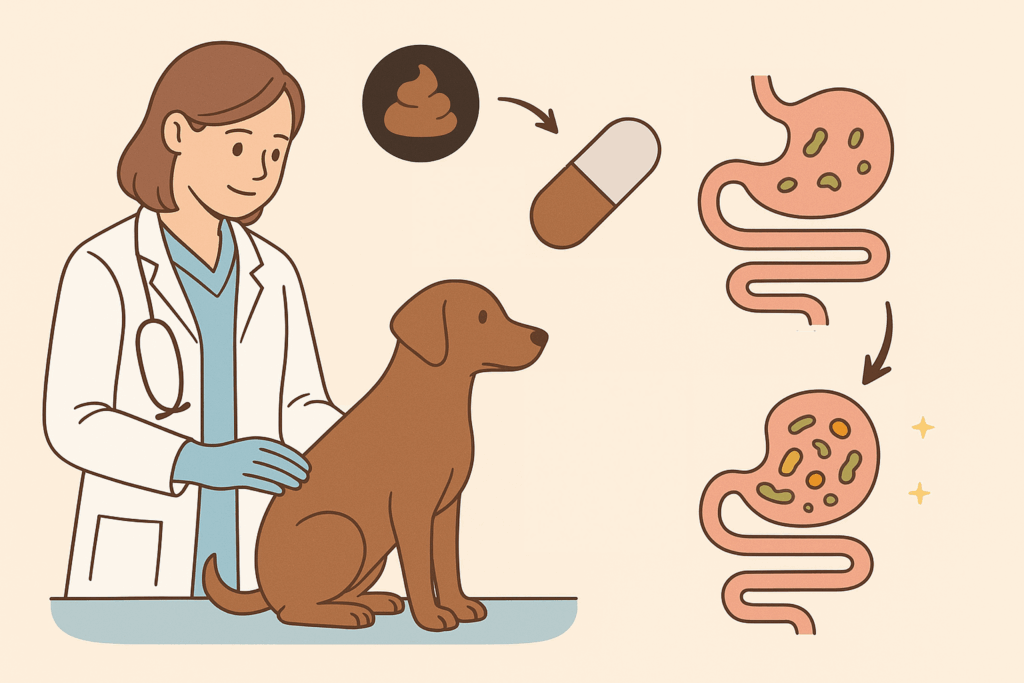
Here at My Pet Nutritionist, we love to look at every avenue when it comes to pet health. There are various support options available for most disease within the body, from drug therapy to dietary tweaks to supplements. One fairly new therapeutic method on offer is Faecal Microbiota Transplant (FMT). A huge number of common diseases in pets are largely down to an unhealthy gut microbiome composition. The gut microbiome is composed of a diverse community of bacteria, fungi, and viruses. The key determinant of gut health lies in the balance among these microorganisms. In a healthy gut, microbial populations exist in a state of equilibrium that supports optimal digestive and immune function. When this balance is disrupted, resulting in an overrepresentation of potentially harmful microbes relative to beneficial ones, the body becomes more susceptible to inflammation, infection, and disease. During FMT treatment, the stool of a healthy donor animal is processed to be safe, and given to the pet with the poor microbiome composition, where it settles in the gut, repopulating the unhealthy gut microbiome into a healthy one. Findings Here There are multiple uses for FMT, so let’s explore these! Uses of Faecal Transplant While FMT is a relatively new support option on the market, it has been shown to have some incredible benefits and uses! Antibiotic resistant infections: the original and intended use of FMT was to help the individual recover from antibiotic resistant bacteria called Clostridioles difficile. Outcompeting the bad bacteria with the good bacteria of a healthy donor animal can help combat this nasty, often recurring infection. IBD/Colitis: research is ongoing into the treatment of IBD and Colitis using FMT. Liver Disease: the treatment of hepatic encephalopathy using FMT is being investigated! A healthy gut means other organs can remain healthy. Autoimmune disease: researchers suggest FMT could play a role in the treatment of autoimmune disease such as arthritis. Metabolic disorders: some metabolic disorders, including poor digestive capabilities and insulin sensitivity may be helped by FMT. Neurological health: as FMT improves gut health, neurological health is directly aided due to the gut-brain axis. Recovery from parvovirus: FMT can be used to repopulate the gut following parvovirus; this is an essential part to recovery, so FMT could be something to discuss with your veterinarian. Findings Here Findings Here Findings Here Production and Administration Many people are curious to know how a FMT sample is made, and how it is administered to the pet. Read on to find out! Prepping the gut to be as healthy as possible using a gut health supplement like our Gut Guardian is an excellent first step. for your pet to get the most out of FMT, the gut needs to be in at least a semi-healthy state. Our analogy is: When you have a premium car with a rusty engine, putting the best oil in won’t necessarily help the car to run better! Due to the nature of FMT, the sample starts life in a healthy donor. Before a sample is taken from the proposed donor, the donor receives a comprehensive examination, and their health history is thoroughly examined. If that donor animal has any history of gastrointestinal issues, they cannot donate faecal matter. Similarly, if they have any ongoing parasitic infection or infectious disease, they will not be able to donate. The use of antibiotics is also not permitted in a donor animal. Once an appropriate donor is selected, a sample of faecal matter is collected, which is then blended into a slurry like liquid with saline solution or any other sterile pure liquid. This solution is filtered, creating a smooth liquid. There are 4 main ways FMT is administered to pets: Oral capsules: the mixture is encapsulated, which allows the pet to swallow the capsule as they would any medication. This method of administration is done while the animal is awake. Enema: using a catheter, the liquid is administered directly via the rectum. This approach is carried out under anaesthetic or sedation. Endoscopy: a tube is inserted into the mouth, and fed through to the gastrointestinal tract. The prepared liquid is pushed through the tube, and deposited into the intestine. This procedure is carried out under anaesthetic. Colonoscopy: a tube is inserted through the rectum and into the gastrointestinal tract, where the prepared liquid is deposited directly into the intestine. This procedure is carried out under anaesthetic. Following the procedure, the pet will be kept at the vet, on strict crate rest to ensure they do not defecate for at least 2 hours. Their condition will be monitored to ensure there are no adverse effects. Probiotics are often recommended as a long term supplement. Findings Here Findings Here How Effective is Faecal Transplant? The big question is: is FMT really that effective? According to studies, FMT appears to be very effective at treating some of the aforementioned health issues! Some small studies suggest a 70-90% improvement in chronic diarrhoea cases after the use of FMT. Those with pets previously suffering with IBD report moderate to significant improvement. In cases where FMT is used as part of a patient’s recovery from parvovirus, it has been noted that recovery tends to be faster, the patient has a better appetite, and stools are firmer, when compared to cases relying purely on conventional treatment. We can also look at the diversity of one’s gut before the transplant and after, which is often done in cases of antibiotic resistance. 1-2 weeks post transplant seems to be the typical length of time for normal healthy gut balance to be restored. What might affect the effectiveness of FMT? Of course, as with any treatment or therapy, there are factors at play that may affect how effective the results of the planned treatment are. Factors which may may FMT less effective include: Quality of the donor sample Quality of screening and filtration of the sample The type of condition being treated, and how severe it is. Method of administration; it is not currently known if the
The eyes are one of a dog or cat’s most important body parts as they give the sense of sight. This sense of sight brings with it an easier ability to socialise and develop mentally. Here at My Pet Nutritionist, we often help those with a variety of eye complaints – nutrition can make a difference to your pet’s eye health! Eye problems in pets can range from minor issues like Conjunctivitis, all the way through to severe conditions like Progressive Retinol Atrophy and other conditions that can end in blindness. This bumper 2-part blog explains what the 9 common eye problems we see most are, how they’re caused, and what you can do to help prevent or improve eye problems in your beloved pet. In Part 2, we will be looking at Glaucoma, Corneal Ulcers, Corneal Dystrophy and Progressive Retinol Atrophy, and we will also discuss some general nutrition and lifestyle tips for eye health! Glaucoma Glaucoma is a condition found commonly in both dogs and cats. When there is a build up of intraocular pressure due to the reduced ability for the eye to drain the fluid, the eye becomes severely painful, and can lead to blindness. Intraocular pressure is the pressure of the fluid located inside the eye; the fluid is called aqueous humor. Typical symptoms of Glaucoma include swelling/bulging of the eye, redness/bloodshot eye, squinting, pawing at the eye, or rubbing it on furniture, increased tear production, and cloudiness. Those with primary Glaucoma have the congenital lack of ability to successfully drain the fluid from the eye, whereas those with secondary Glaucoma do so as a result of trauma or other eye issues. Breeds most affected by primary Glaucoma include the Cocker Spaniel, Basset Hound, Shar Pei, American Cocker Spaniel, Beagle and Husky. In most cases, the vet will have a rough idea as to what the problem with your pet’s eye is just form looking at it, however to confirm their suspicions, they will use a handheld device called a Tonometer, which measures the pressure inside the eye. Once diagnosed, to give the pet the highest chance of avoiding vision loss, they will act immediately with a treatment plan. Treatment could include surgery, eye drops, or even oral medications depending on the severity. Always accept offers of painkillers for pet as this condition is painful! Oral medications to suppress aqueous humor production are more commonly used now than ever! Usually the chance of recovery heavily depends on the type of Glaucoma and speed of diagnosis and treatment. Those with primary Glaucoma tend to go blind in the vast majority of cases, whereas those with secondary Glaucoma tend to stand more chance of recovery if the right treatment is given quickly after symptoms start. Findings Here Findings Here Corneal Ulcer Corneal Ulcers are one of the most common eye complaints we see. Characterized by their dented appearance, ulcers can happen for a number of reasons, and can be of various levels of severity. These ulcers are wounds on the front of the eye, and they can be shallow or deep. The deeper the wound, the more damage done, and the harder they are to treat. Common symptoms in those with eye ulcers include squinting, increased tear production, cloudiness, discharge in the corners of the eye, rubbing the eye with a paw or against furniture, and reduced awareness of the pet’s surroundings. Ulcers are most common in dogs and cats with bulging eyes (typically brachycephalic breeds like French Bulldogs, Pugs, Persians and Exotic Shorthairs) due to their vulnerability to eye trauma, but can occur in any breed! During diagnosis, your veterinarian will use a dye known as a fluorescein stain, which is dropped into the eye. This stain will show any dents or imperfections in the cornea, allowing a diagnosis of the ulcer. There are multiple types of ulcer, and the treatment recommended will heavily depend on the type of ulcer at hand. In the majority of cases, an antibiotic eye drop will be prescribed to prevent any infection building in the wound, as well as a lubricating eye drop to aid smoothness while the eye is injured. All cases will be prescribed a pain killer medication which is essential for your pet’s comfort. You will also be instructed to not allow your pet to scratch the eye/face area. More severe cases will require surgery, the most common surgery being a Diamon Burr Debridement whereby the dead tissues in the eye are removed. Top Tip: ask your vet to make a blood serum! For mild to moderate ulcers, this serum can be incredibly affective in quickly treating the ulcer! In some severe cases infection sets in, and begins eating away at the cornea, further increasing the risk of blindness, and spreading of infection. This type of ulcer is known as a melting ulcer and will require immediate action. The eye will begin to have a mushy appearance in these cases. Findings Here Findings Here Findings Here Corneal Dystrophy Another eye complaint characterised by cloudiness of the lens, Corneal Dystrophy is a hereditary condition whereby abnormal deposits can be found on the cornea. There are not one, but three types of Corneal Dystrophy our pets can face. The three types are: Epithelial: mildly painful, causing severe light sensitivity and squinting. Deposits form on the outermost layer of the cornea. Stromal: usually painless with mild visible changes to the eye such as hazy spots. Deposits of fats and minerals form on the middle layer of the cornea. Endothelial: very painful due to swelling within the eye. Affects the innermost layer of the cornea. Can result in ulcers. The symptoms for this condition are usually very obvious to the naked eye and include cloudiness of the lens, blueish, grey or white patches on the cornea, brown or black spots in the iris, light sensitivity, bloodshot eyes and redness of the tissues surrounding the eye, and in the most severe cases, blindness! Diagnosis is usually quick and simple, using handheld
The eyes are one of a dog or cat’s most important body parts as they give the sense of sight. This sense of sight brings with it an easier ability to socialise and develop mentally. Here at My Pet Nutritionist, we often help those with a variety of eye complaints – nutrition can make a difference to your pet’s eye health! Eye problems in pets can range from minor issues like Conjunctivitis, all the way through to severe conditions like Progressive Retinol Atrophy and other conditions that can end in blindness. This bumper 2-part blog explains what the 9 common eye problems we see most are, how they’re caused, and what you can do to help prevent or improve eye problems in your beloved pet. In Part 1, we will be looking at Cataracts, Conjunctivitis, Dry Eye, Cherry Eye and Entropion. Cataracts With their classic ‘cloudy eye’ appearance, cataracts are one of the most commonly diagnosed eye issues in pets, especially in dogs. Cataracts occur when the lens of the eye becomes clouded, and vision becomes impaired. There are 3 types of cataract, each with differing levels of severity and cloudiness: Incipient: an extremely small, pinpoint-like opacity in the lens. This type of cataract does not require treatment, and doesn’t generally cause any form of vision impairment. Immature: partial lens opacity. Starts affecting vision, and is usually treatable. Mature: full lens opacity. Vision is lost, and treatment may be possible. Cataracts are typically an age related condition, and is far more prevalent in the older population of pets than the younger population. The prevalence of cataracts is high in dogs older than 4-7 years (breed dependent), and very likely in those ages 10-13 or older. Breed predisposition is also a determining factor for cataracts. Some studies suggest that cataracts are most common in 60 breeds of pure bred dog as opposed to mixed breed dogs. Common breeds that tend to be prone to cataracts include the Miniature Poodle, Miniature Schnauzer, Smooth Fox Terrier and Boston Terrier. In a lot of cases, hereditary genetics are a determining factor too! Quite often if a parent pet has cataracts, it’s offspring will develop them too. These often affect dogs and cats earlier in life, however can appear later in life too. Dogs with Diabetes Mellitus are largely at risk of cataracts; in fact, within their first year of diagnosis, around 75-80% of dogs with diabetes develop cataracts. Trauma and inflammation are also associated with cataracts. In most cases, cataracts are treated surgically. This surgery entails the clouded lens being removed (high frequency ultrasound breaks up the clouded material, and this is then suctioned out), and an artificial lens being inserted. This treatment is quite costly, but does greatly improve the lives of those who receive it. Minor cases of cataracts are treated with corticosteroid or non-steroidal eye drops which manage inflammation in and around the eye. Findings Here Findings Here Conjunctivitis Conjunctivitis is the most common eye infection in pets. The swelling of the pink tissue inside the eye cavity becomes inflamed, which causes the outward symptoms associated with conjunctivitis. There are many causes of conjunctivitis, such as: Bacterial, viral or fungal infection: these all cause inward stress leading to outward stress, and the pathogens themselves cause inflammation. Allergies: dust, dander and pollen can cause conjunctivitis. Foreign bodies: grass seeds are a big one! Grass seeds are nasty – they migrate inside the pet! The eyes are somewhere these seeds can get stuck. The inflammation caused by foreign bodies can cause conjunctivitis. Dry eye: when the tear glands are underactive, the eye lid becomes abrasive which causes inflammation around the eye. Read more on this in the next section! Breed predisposition: some breeds, namely those with unusually shaped eyes or who are susceptible to blocked tear ducts such as Pugs, French and English Bulldogs, Clumber Spaniels, Great Danes are predisposed to having conjunctivitis regularly. Environmental Irritants: areas of pollution outside, including smoke, gardening products etc can irritate the tissues around the eyes, causing conjunctivitis. The signs to look out for which would commonly be associated with conjunctivitis include redness in the eye cavity, bloodshot eyes, discharge from the eye, squinting or rubbing the eye on furniture or with their paw, and unusually matted fur around the eye. Treatment is usually done at home using prescription treatments from the vet, such as antibiotic eye drops, steroids or antihistamines, and massage of the tear drops. Saline flushes may be carried out at the vet in severe cases. Hypochlorous Acid products such as Leucillin may be beneficial for your pet, and chamomile tea soaks are often very soothing too! Findings Here Findings Here Dry Eye Known clinically as Keratoconjunctivitis sicca (KCS), Dry Eye happens when the pet has an underactive tear gland. The role of the tear glands is to lubricate the eye, so when these are not producing enough tears, they eye becomes dry. This leads to inflammation of the tissues in the eye cavity, and sticky discharge, and eventually blindness if not treated. In order to diagnose Dry Eye, the Schirmer Tear Test will be performed during which the veterinarian will place a specific paper strip under the lower eyelid for 60 seconds. The results are interpreted by identifying how far up the strip the tears have absorbed. There are various causes of Dry Eye such as: Chronic infection: severe or long term eye infections can cause dry eye. Congenital issues: pets may be born without tear ducts. Autoimmunity: the body’s immune system attacks itself; in this case, it attacks the tear glands. Neural issues: some pets may have problems with the nerves that stimulate tear production by tear glands. Trauma: injury to the eye can cause the glands to stop working Medications: unfortunately all medications come with side affects. Some medications hold the side affect of reduced tear production, which can then lead to dry eye. Treatment for dry eye is typically treated at home with a combination of medications to promote
Here at My Pet Nutritionist, one of the topics we get asked about a lot is Addison’s Disease. This is something that can be heavily influenced by diet and lifestyle, so this blog post is a great read for those with a dog suffering from this awful and debilitating disease. Worldwide, somewhere between 1in 500 and 1 in 1000 dogs is affected by Addison’s Disease. Closer to the home of My Pet Nutritionist, a study was carried out in 2016 to determine the estimated number of dogs with Addison’s Disease in the UK. It was estimated under 1% of the dog population have it, though this may have changed as time has passed. Most commonly, young female dogs tend to be most prone to Addison’s Disease. Severe cases of Addison’s Disease lead to Addisonian Crisis, which is life threatening. Read on to find out more about this nasty disease! Findings Here What is Addisons Disease? The clinical name for Addison’s Disease is Hypoadrenocorticism. This is an endocrine disease; a disease affecting the hormone system. Addison’s specifically involves the Adrenal glands which are located near the kidneys. These glands produce lots of extremely important hormones controlling various health factors, including the control of metabolism, stabilisation of blood pressure, and keeping blood sugar levels steady. It is also involved in the immune system and part of the stress response. The adrenal glands, of which there are two, are comprised of two parts each; the inner medulla and the outer cortex. The inner medulla produces adrenaline, and the outer cortex produces two hormones called cortisol and aldosterone. Addison’s Disease concerns the outer cortex. Those suffering with the disease do not produce sufficient amounts of cortisol or aldosterone. Findings Here Symptoms and Causes There are a range of symptoms seen in those with Addison’s Disease. Typically you will see the following symptoms in mild to moderate cases: Letharghy Vomiting Diarrhoea Weight loss Loss of appetite Low mood Muscle weakness Increased thirst Increased urination In those entering Addisonian Crisis, a smaller range of more severe symptoms are possible, including: Severe dehydration (due to the inability for the body to control the salt and water balance in tissues) Irregular heart rate and blood pressure (due to the lack of aldosterone which is responsible for helping the kidneys retain sodium and excrete potassium; a crucial part of stable blood pressure) Collapse Cold extremities Persistent and severe vomiting and diarrhoea The milder symptoms are often preventable and manageable with the right lifestyle, diet and medication regimes, however those experiencing one or more of the more severe symptoms must seek veterinary aid immediately as they may be entering Addisonian Crisis which is life threatening. There are multiple potential causes for the onset of Addison’s Disease. Let’s start with autoimmunity. Autoimmune diseases happen when the body’s immune system attacks itself and other parts of the body. When a dog has an autoimmune disease affecting the adrenal glands, the body attacks the glands which damages them, causing a lack of the associated hormones. There is a specific type of Addison’s Disease known as Latrogenic Addisons. This is caused by the overuse of pharmaceuticals when a dog is treated for Cushing’s Disease (which is due to overactive adrenal glands). The drug used to treat Cushing’s Disease suppresses adrenal gland activity to better control the amount of hormones it produces, however when this drug suppresses the glands too far, Addison’s Disease is a huge risk as the glands will go from overproducing to underproducing the necessary hormones. Addison’s Disease can also be a secondary disease. The Pituitary Gland and the Adrenal Glands are part of the HPA Axis (Hypothalamic-Pituitary-Adrenal). The Adrenal Glands are largely controlled by the Pituitary Gland, so if there is a defect on the Pituitary such as a tumour or other issue, this can massively affect the functioning of the Adrenal Glands, causing partial or complete failure to produce Cortisol and Aldosterone. Another form of secondary Addison’s Disease is due to the sudden stopping of long-term steroid treatment. The shock of this on the body can cause the Adrenal Glands to stop working properly. Genetic predisposition also plays a role in the cause of Addison’s Disease. While any breed of dog can be affected, some breeds are more prone to Addison’s than others. Breeds that are genetically predisposed include Portuguese Water Dogs, Great Danes, West Highland White Terriers, Standard Poodles, Bearded Collies, Rottweilers, Leonbergers, Labradors, Soft Coated Wheaten Terriers and Nova Scotia Duck Tolling Retrievers. Findings Here Findings Here Findings Here Diagnosis If your dog displays any of the aforementioned symptoms, it is important you consult your vet who will run the appropriate diagnostic tests. Firstly, the vet will run a full blood panel, and also a urinalysis to detect any possible imbalances such as low sodium and/or high potassium which would pinpoint an issue with adrenal function. Dilute urine would also be a sign of potential adrenal function loss, but could also be a sign of diabetes insipidus. The next test performed, which confirms Addison’s Disease, is called the ACTH Stimulation Test. During this test, a blood sample is taken, before the dog is given a synthetic ACTH-like drug, intravenously. Two hours after the injection is given, another blood sample is taken. Cortisol levels in both blood samples are compared with one another, to check how much cortisol the body has produced in the presence of the ATCH-like drug (which mimics the Adrenocorticotropic Hormone secreted by the Pituitary Gland). Your veterinarian will then interpret these results. This is the same test used to diagnose Cushing’s Disease. It is important for both you and your veterinarian to note that if your dog has been on steroidal drugs before the ACTH test is performed, a false positive for Addison’s Disease is possible. Findings Here Conventional Treatment Treatment is absolutely necessary in those with Addison’s Disease; if it is not kept on top of, it can become life threatening! Initially, your vet may recommend your dog stays in the vet practice
Intolerances are one of the hottest topics here at My Pet Nutritionist! We see a huge number of itchy dogs, many with intolerances and general inflammation. Inflammation can cause many more health problem in both us and our pets, but what causes it? It can be very tricky to work out the underlying cause of inflammation, so reviewing the diet may be a great first step! In this blog post, we look at the difference between allergies and intolerances, and their link to inflammation in the body. Intolerance vs Allergy The terms ‘intolerance’ and ‘allergy’ are not interchangeable – something many pet owners don’t realise! While both conditions have similar symptoms, the clinical mechanism of disease is very different! One is curable, the other is not. Let’s take a look at each in turn. Intolerances are a histamine response to a stimulus caused by poor gut health. Intolerances can be helped or even cured through the use of gut health supplements such as our popular supplement Gut Guardian. When the gut integrity is compromised, food particles leak from the gut and into the bloodstream where they are detected and recognised as foreign bodies. When the body recognises a foreign body it will either release a specific antibody (in the presence of a chemical compound called histamine which helps the immune system fight infection, and causes the typical reaction symptoms you see in those with an allergy or intolerance. Healing the gut goes a long way in the reduction of intolerances, and often in the cure! Allergies are not curable. These are caused by a malfunction of the immune system; whereby the allergen causes severe reactions. True allergic reactions range from skin rashes and digestive issues to anaphylactic shock. Allergies can cause death, whereas intolerances cannot. Once an allergen is discovered, it is important not to feed this as part of the pet’s diet. Findings Here What is Inflammation? In order to understand the link between intolerances and inflammation, it is important to know exactly what inflammation in the body really is, and where it can happen. Inflammation is part of the body’s immune response. It can happen following trauma, during infection, due to an autoimmune condition whereby the immune system attacks the itself, due to exposure to toxins and hazardous substances like flea treatments, and also in the presence of a foreign body, be it food particles leaking from an unstable gut wall, or a physical object inside the pet. Outwardly, it is characterised by swelling, redness, heat, pain when touched, and even loss of use of the body part it is present on. When inflammation occurs outwardly, it is easy for us to see it, and help the pet, however when it is internal inflammation, it’s not visible to the naked eye which makes it far trickier to deal with! In the presence of a trigger for inflammation, specialised molecules are released by the immune system, which causes an increase in blood flow to the affected area, which causes the blood vessels to leak. This increased blood flow teamed with the leaky blood vessels is the transport system for immune cells to reach the area in an attempt to fix the problem. In pets with intolerances, we often deal with internal chronic inflammation which is more long term, and this constant rush of immune cells to these areas can cause deterioration in previously healthy tissues. Inflammation in the body is not a good thing – it’s something we want to aim to reduce or keep at bay entirely. Inflammation can cause major pain, gastrointestinal, digestive and heart issues, and can also reduce the mental health of the pet, as well as cognitive capabilities. Many pet owners mistakenly assume inflammation is always visible, and can only happen on the outside of the body. Internally, inflammation can occur throughout the body! From brain to gut, inflammation can happen. When it comes to food intolerances, the direct link with inflammation is via the gut, however this then impacts other parts of the body, which we will discuss next! Findings Here The Link Between Intolerances and Inflammation So, is there a link between intolerances and inflammation? The short answer is ‘yes’! Let’s take a deeper dive into this link. As we have previously mentioned, symptoms often associated with inflammation can vary massively both internally and externally. In terms of inflammation, the outward signs of inflammation are: Itching Skin redness Hair loss Chronic ear infections (these are caused by intense swelling in the inner ear) Visible swelling Vomiting Diarrhoea Food intolerances can be traumatic for the body, which means inflammation occurs throughout the body, starting from the gut. Due to the series of gut axes (including the gut-brain axis, the gut-joint axis and the gut-skin axis), inflammation in the gut, indirectly causes inflammation in these other parts of the body, so a pet with intolerances can truly have their whole body affected! When the body detects what it thinks are ‘foreign bodies’ in the form of leaked food particles, the immune system send lots of histamines to the area, which means their transport system (blood) rushes to the affected area quickly, causing the inflammation. The knock on effect of this inflammation in the gut is seen as trauma in other body parts, and so the immune system rushes more blood to these areas too, which again creates inflammation. The initial inflammation, or direct inflammation, caused by food intolerances is called mucosal inflammation, which is inflammation of the mucosal lining of the digestive tract. Findings Here Findings Here How Can We Reduce Inflammation Caused By Food Intolerances? There are a number of things we can do to help reduce inflammation due to food intolerances, but ultimately helping the root cause will be the most beneficial pathway. You will find a link to our consultations at the end of this blog post; booking one of these would be incredibly beneficial for you and your pet if they suffer with intolerances you can’t get control of.