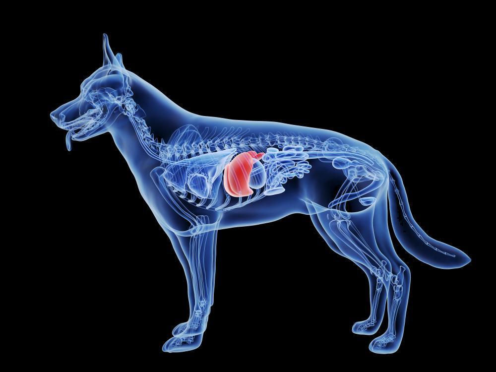
As our pets eat their food, it passes from the stomach and into the small intestine. Whilst some of the digestive processes have already started, most of the chemical digestion that occurs in the small intestine relies on the activities of the pancreas, liver, and gallbladder. Let’s take a closer look at the function of the pancreas and its role in our pet’s digestion. What is the Pancreas? The pancreas is a gland found in the digestive system of our pet. It is made up of a head, a body and a pointy tail-like end. It is in the upper abdomen behind the stomach and carries out two main roles in the body: The pancreas produces: Enzymes that break down foods in the intestine Hormones that regulate blood sugar levels The pancreas is made up of small clusters of glandular epithelial cells. About 99% of these clusters constitute the exocrine portion of the organ. These cells secrete a mixture of fluid and digestive enzymes known as pancreatic juice. Pancreatic juice consists mostly of water, but it also contains some salts, sodium bicarbonate and several enzymes. The sodium bicarbonate gives the pancreatic juice a slight alkalinity that buffers the stomach acid in the chyme that has just arrived in the small intestine. It also stops the action of pepsin and creates the correct pH for the action of the digestive enzymes to function. The digestive juices that are made in the pancreas flow into the small intestine through a tube known as the pancreatic duct. In most bodies, this duct is joined by a similar duct coming from the gallbladder (the bile duct) before it reaches the small intestine. There is a circular muscle (sphincter) at the shared opening of the two ducts. This muscle controls the release of the digestive juices into the small intestine. The digestive juices usually only start working once they enter the small intestine. But if the pancreas is inflamed (pancreatitis), they already become active in the pancreas. This can start causing a range of issues. Digestive Enzymes Enzymes are biological molecules (typically proteins) that significantly speed up the rate of virtually all of the chemical reactions that take place within cells. When discussing the pancreas, there are three main types of enzymes: Lipases to break down fats Proteases to break down proteins Amylases to break down starch The remaining 1% of the clusters called pancreatic islets (islets of Langerhans) form the endocrine portion of the pancreas. These cells secrete the hormones glucagon, insulin and more. These hormones usually help to regulate blood glucose levels, stopping them from getting too high or too low. Glucose Regulation Glucose is a 6-carbon structure with the chemical formula C6H12O6. It is a source of energy for every organism in the world and is essential to fuel both aerobic and anaerobic cellular respiration. Glucose often enters the body in isometric forms such as galactose and fructose (monosaccharides), lactose and sucrose (disaccharides), or starch (polysaccharide). The body stores excess glucose as glycogen, which becomes liberated in times of fasting. Glucose is also derivable from products of fat and protein break-down through the process of gluconeogenesis. Once glucose is in the body, it travels through the blood and to energy-requiring tissues. There, glucose is broken down in a series of biochemical reactions releasing energy in the form of ATP. The ATP derived from these processes is used to fuel virtually every energy-requiring process in the body. As glucose is so important to life, it stands to reason that regulation of it is incredibly tight. And as we have noted, there are a number of hormones involved in this process. Hormones involved in Glucose Regulation Hormones Involved: Insulin is a peptide hormone. Insulin plays an important role to keep plasma glucose value within a relatively narrow range throughout the day. Insulin’s main actions are: (1) In the liver, insulin promotes glycolysis and storage of glucose as glycogen (glycogenesis), as well as conversion of glucose to triglycerides (2) In muscle, insulin promotes the uptake of glucose and its storage as glycogen (3) in adipose tissue, insulin promotes uptake of glucose and its conversion to triglycerides for storage Insulin lowers glucose levels. Glucagon: Glucagon acts exclusively on the liver to antagonise insulin effects on hepatocytes. It enhances glycogenolysis and gluconeogenesis. It also promotes oxidation of fat, which can lead to the formation of ketone bodies. Glucagon increases glucose levels. Levels of both insulin and glucagon vary depending on nutrient intake. The Fed State: The fed state occurs after a meal and is also known as the absorptive state. It is characterised by a high insulin to glucagon ratio. Anabolic metabolism dominates in the fed state largely to replenish fuel stores, this is achieved by glycogen synthesis, fatty acid synthesis and protein and amino acid metabolism. The Fasting State: The fasting state occurs between meals and ensures a maintenance of blood glucose level. This state is characterised by a low insulin to glucagon ratio. This low insulin to glucagon ratio overall promotes catabolism in comparison to the fed state. In this state the major pathways include gluconeogenesis, glycogenolysis, protein catabolism, lipolysis, and ketone body metabolism Endocrine cells secrete these respective hormones in response to external signals, such as nutrient intake or stress, via humoral, neural or hormonal signalling pathways. The Brain-Islet Axis The pancreas is highly innervated with both parasympathetic and sympathetic nerve fibres from the autonomic nervous system. At the same time, insulin receptors are widely distributed within the brain. In rat studies, lesions in various brain regions were shown to affect pancreatic hormone secretion. Norepinephrine also inhibits insulin secretion, which is an important aspect of the fight-or-flight response. Insulin release is stimulated by the cephalic phase, which is the period of anticipating a meal, to prepare the body to adequately respond to incoming nutrients. The Liver-Islet Axis The liver has a key role in glucose homeostasis by storing (glycogenesis) or releasing (glycogenolysis/gluconeogenesis) glucose. Liver Guard The Gut-Islet Axis The gut releases various
When we talk about inflammation, we generally see it as something bad and something we need to get rid of. But like all things ours and our pet’s body does, it serves a purpose. Despite this, there is a difference between acute and chronic inflammation. So, let’s get to grips with the purpose of inflammation, what causes it and when it becomes problematic. We’ll also take a look at some nutrition tips to help modulate this response. What is Inflammation? Inflammation is part of the body’s defence mechanism. It is the process by which the immune system recognises and removes harmful and foreign stimuli and begins the healing process. The immune system senses something is wrong and sends its soldier immune cells to tackle the issue, which is why the hallmark signs of inflammation; heat, redness, swelling and pain occur. Inflammation can be either acute or chronic. Acute Inflammation Tissue damage due to trauma, microbial invasion, or noxious compounds can induce acute inflammation. It starts rapidly, becomes severe in a short time and symptoms may last for a few days, an example is bacterial infection. Chronic Inflammation Chronic inflammation is also referred to as slow, long-term inflammation lasting for prolonged periods of several months to years. Generally, the extent and effects of chronic inflammation vary with the cause of the injury and the ability of the body to repair and overcome the damage. For the most part, acute inflammation serves a purpose, and the issue is resolved. Chronic inflammation is what becomes the issue. Chronic inflammation can be a result of the following: Failure of eliminating the agent causing an acute inflammation like a recurrent infection or exposure to an allergen. Exposure to a low level of a particular irritant or foreign material that cannot be eliminated by enzymatic breakdown or phagocytosis in the body. An autoimmune disorder in which the immune system recognizes the normal component of the body as a foreign antigen and attacks healthy tissue. A defect in the cells responsible for mediating inflammation leading to persistent or recurrent inflammation. Inflammatory and biochemical inducers resulting in oxidative stress and mitochondrial dysfunction such as increased production of free radical molecules, advanced glycation end products (AGEs), uric acid (urate) crystals, oxidized lipoproteins, and homocysteine. Factors that may increase the risk of chronic inflammation include: Ageing Increasing age is associated with elevated levels of inflammatory markers – this could be due to mitochondrial dysfunction or free radical accumulation over time. Obesity Fat tissue is often described as an endocrine organ. It secretes multiple adipokines and inflammatory mediators. There is increasing data showing that the adiposity correlates with levels of pro- inflammatory markers. Diets high in saturated fats This is increased further in those who are obese. Low sex hormones Data has indicated that testosterone and oestrogen can suppress the production of pro-inflammatory markers – so when there are low levels (in the case of neutered pets) inflammation can increase. Findings Here Stress Physical and emotional stress is associated with inflammatory cytokine release – which is why the modulator of the stress response, cortisol is deemed anti-inflammatory. However, prolonged stress can result in cortisol dysfunction. It is thought that cortisol binding is downregulated, and it fails to function. In humans, stress-induced inflammation has been linked to a range of chronic conditions like osteoporosis, myopathy, sciatica and more. Findings Here Poor sleep/recovery Again, cortisol plays a role here. There is a natural diurnal cortisol in dogs. You will see a natural rise in cortisol in the morning, with it progressively dropping throughout the day. Dogs are similar to humans in this respect. If sleep is disrupted or our dogs are unable to follow their diurnal rhythms, cortisol levels can become dysfunctional. Study after study have highlighted that poor sleep is associated with higher levels of inflammatory biomarkers. Findings Here The Importance of Sleep Dietary sensitivities/allergens What goes on in the gut doesn’t always stay in the gut and translocation can occur across the gut wall. This means that particles end up in places they shouldn’t, calling the immune system to action. 7 steps to Optimal Gut Health in Pets Signs of Chronic Inflammation: Body pain Anxiety/depression Gastrointestinal issues like constipation, diarrhoea, and acid reflux Unintended weight gain/loss Frequent infections It is crucial to understand the driver of chronic inflammation, and assessment from a qualified practitioner will be necessary here. But in managing chronic inflammation there are some beneficial changes that can be made: Top Tips: limit intake of ultra-processed foods ensure an appropriate body weight Obesity in Pets Part One Part Two – high intake of dietary fibre is associated with lower inflammatory markers – increase intake of fruits and vegetables – blueberries, apples, brussels sprouts, and broccoli are all high in natural antioxidants and polyphenols which may help protect against inflammation. Does My Dog Need Antioxidants? – Curcumin – significant improvements are noted in inflammatory models in animals when administered turmeric. – Omega-3s – increased intake of omega-3 is associated with lower levels of many inflammatory markers. Essential Fats For My Dog’s Diet – Micronutrients – magnesium, vitamin D, vitamin E, zinc and selenium levels are all associated with inflammatory markers. They all exert inflammatory modulating effects in the body. The Importance of Vitamin D for Cats and Dogs Why Does My Dog Need Minerals? Why Zinc is Important for Your Dog Acute inflammation is necessary, and it is a process by which the body returns to homeostasis. The issue is when inflammation becomes chronic, and we know there are several reasons why this may occur. It’s essential to understand the contributing factors to chronic inflammation, but there are lifestyle changes that can help. Check out our services if you’d like to see how we can help. Thanks for reading, MPN Team
Epilepsy is the most common chronic neurological disorder in dogs, with a formerly reported prevalence of between 0.5% and 5% in the canine population. Epilepsy is not one single disease process but can be elicited by multiple causes and can be classified as genetic (primary or idiopathic), structural and of unknown origin/etiology. Let’s get to grips with what it is and some of the causes. What is Epilepsy? Epilepsy occurs naturally in many species including rodents, cats, dogs, horses, cattle, goats, non-human and human primates. It is the most common acquired chronic neurological disorder in humans having a a worldwide incidence of approximately 50–100 cases per 100,000 persons (higher in undeveloped countries) and a prevalence of 4–10 per 1000 persons. In humans there are over forty epileptic syndromes and related conditions. In dogs, however, epilepsy is not usually differentiated into syndromes. Most dogs with recurrent seizures have no identifiable underlying cause and are classified as having idiopathic epilepsy. The lack of canine epilepsy classification may be due to the difficulty of seizure description and classification, and partly because electroencephalography (EEG) is not routinely used in veterinary neurology clinics. To that end, we simply define epilepsy as a chronic neurological condition characterised by recurrent epileptic seizures. Idiopathic epilepsy in dogs Most dogs with recurrent seizures are thought to have idiopathic epilepsy, in short, no underlying cause of the seizure can be identified. In these cases the first seizure usually occurs between 6 months and 6 years of age, but occasionally seizures have been known to start as young as 3 months and as late as 10 years of age. There appears to be a hereditary basis for idiopathic epilepsy, with several breeds being affected: Beagles German Shepherds Labrador Retrievers Golden Retrievers Bernese Mountain Dog Viszlas Keeshonds English Springer Spaniel Recently a mutation found on the Epm2b gene has shown to be causal in miniature wire-haired dachshunds. The Theory of Epilepsy The pathophysiology of epilepsy is often suggested to be an imbalance between excitation and inhibition in neurotransmitters; increased excitation or decreased inhibition may lead to epileptiform activity in the brain. It is considered that there is a fine balance between the excitatory glutamate neurotransmitter and the inhibitory GABA neurotransmitter. This theory has been tested in dogs and researchers found significantly less GABA and more glutamate in cerebral spinal fluid (CSF) from epileptic patients when compared to normal controls. Sadly, there appears to be a double-edged sword too, many receptors in the brain undergo altered expression following seizures and this may lead to changes in excitability of the brain and be involved in further pathogenesis of seizure disorders. Temporal Lobe Epilepsy In Dogs Temporal lobe epilepsy is the most common partial seizure disorder in adult humans and there are reports of it occurring in dogs. It has also been suggested that “fly-biting” or “fly-catching”, a behaviour in which dogs snap aimlessly into the air as if trying to catch a fly, may have a temporal lobe origin. Treatment Since epilepsy is often associated with inhibition and excitation in the brain, antiepileptic drugs (AEDs) are used to alter the excitability of the brain and aim to reset the balance. There are many mechanisms thought to be involved in the action of antiepileptic drugs. They may functionally block voltage-gated sodium channels They may directly or indirectly enhance inhibitory GABAergic transmission They can inhibit excitatory glutamatergic neurotransmission They can modulate calcium ion channels Vagus Nerve Stimulation The method used in humans was devised in canine models and has since been used with mixed results. It is based on the idea that repetitive electrical stimulation of the canine cervical vagus nerve interrupts or abolishes motor seizures. Findings Here Epilepsy and Diet The Keto Diet The ketogenic diet—a high‐fat, low carbohydrate, and moderate protein protocol—has been used to treat epilepsy for nearly 100 years in both children and adults. A randomised controlled trial on childhood epilepsy showed promising results with 38 and 7 % of children on KD diets having >50 and 90 % seizure reduction, respectively. In comparison, only 6 % of the children on control diets achieved >50 % seizure reduction, with no children achieving >90 % seizure reduction. Findings Here It therefore makes sense that a ketogenic diet has been considered for use in dogs. One study of 21 dogs found that seizures were reduced significantly in dogs fed a proprietary ketogenic diet for 3 months. No improvement was seen in dogs fed a standard diet for the same duration. For 3 subjects, seizures appeared to stop entirely, demonstrating a 100% reduction in seizure frequency. In 7 dogs, seizures decreased by at least 50%, and another 5 dogs experienced a lower seizure frequency overall. Findings Here A Whistle Stop Tour of Keto A Keto Diet for Pet Cancer Omega-3 Supplementation Omega-3 fatty acid deficiency has an interesting role in seizure outcome. It is thought that Omega-3 fatty acids can enhance GABAergic transmission in animals with epilepsy by stimulating the formation of new hippocampal interneurons or by altering expression of calcium-binding proteins. When trialled in a patient with drug-resistant epilepsy, seizures reduced by 85%. Findings Here Essential Fats For My Dog’s Diet Epilepsy and Allergy In humans there are a number of reports that associate allergy with epilepsy. For example, children with allergic symptoms have a 76% increased subsequent risk of epilepsy. It has been found that in these individuals, hypoallergenic diets can reduce the frequency of seizures. Again, we must consider that this may apply to dogs. One study followed dogs with allergic disease. It included eight refractory epileptic dogs and seven were found to have gastrointestinal or skin allergies in conjunction with their refractory seizures. Introduction of an exclusion diet reduced seizures to an “acceptable level” in seven out of eight dogs. Behavioural abnormalities associated with seizures were eliminated in all cases. Findings Here Elimination Diets For Dogs Epileptogenic Toxins Many researchers posit that it’s not only certain foods that are epileptogenic, but toxins can also be problematic. Whilst we’re not talking
There are two branches of science that help us understand both ours and our pet’s bodies. Anatomy is the science of body structures and the relationships amongst them. We learned about anatomy through dissection – we carefully cut up body structures to see what they looked like and the relationship they had with those around them. Physiology is the science of body functions -in short how those body parts and structures work. Both branches have sub-branches, for example we may be interested in pathological anatomy – the structural changes associated with disease. In this blog, we are going to take a look at renal physiology – in short, the function of the kidneys. The Urinary System The kidneys form part of the urinary system and overall, this system consists of: Two kidneys Two ureters One urinary bladder One urethra As body cells carry out metabolic activities they consume oxygen and nutrients. During this process, waste products are made. These waste products must be eliminated from the body because if they are left to accumulate, they can become toxic. Just like the respiratory system eliminates carbon dioxide, the urinary system picks up these waste products. But this is not the only function of the urinary system. Functions of the Kidneys Excretion of wastes: By forming urine, the kidneys help excrete waste from the body. Some of these are a result of metabolic processes, like urea and ammonia, which is as a result of the deamination of amino acids, but it also includes creatinine which is a result of breaking down creatine phosphate. Finally, it includes uric acid from the catabolism of nucleic acids. These compounds are all known as nitrogenous wastes as they are wastes that contain nitrogen. The remainder of wastes are foreign substances that have entered the body, like medications and environmental toxins. This is why we look at kidney health when considering the toxins our dogs must break down. Does My Pet Need to Detox? Regulation of blood composition: The kidneys help regulate the blood levels of several ions including sodium, potassium, calcium, chloride, and phosphate. This is achieved by controlling the excretion of said ions into urine. Why Does My Dog Need Minerals? Regulation of blood pH: The kidneys excrete a variable amount of hydrogen ions into the urine and conserve bicarbonate ions which are an important buffer of hydrogen in the blood. Both of these activities help regulate blood pH. Regulation of blood volume: The kidneys adjust blood volume by conserving or eliminating water in the urine. An increase in blood volume increases blood pressure and a decrease in blood volume decreases blood pressure. Regulation of blood pressure: The kidneys secrete the enzyme renin which activates the renin-angiotensin-aldosterone pathway. Increased renin increases blood pressure. Production of hormones: The kidneys produce two hormones, calcitriol helps regulate calcium homeostasis and erythropoietin stimulates the production of red blood cells. Regulation of blood glucose level: Like the liver, the kidneys can use certain amino acids like glutamine in gluconeogenesis, which is the production of new glucose molecules. They can then release glucose into the blood to help maintain a normal blood glucose level. The Contributions of The Urinary System for All Body Systems Skeletal System The kidneys help adjust levels of blood calcium and phosphates needed for building extracellular bone matrix. Nutrition for Bone and Joint Health Muscular System The kidneys help adjust calcium levels for contraction of muscle. Nervous System Through the process of gluconeogenesis, the kidneys provide glucose for ATP production in neurons, especially during fasting or starvation. Endocrine System The kidneys participate in the synthesis of calcitriol, the active form of Vitamin D. They also release erythropoietin which is the hormone that stimulates the production of red blood cells. Cardiovascular System As noted previously, the kidneys play a key role in the regulation of blood volume, pressure, and composition. Lymphatic System and Immune Function By increasing or decreasing their reabsorption of water filtered from blood, kidneys help adjust volume of interstitial fluid and lymph. Urine also flushes microbes out of the urethra. Respiratory System The kidneys and lungs cooperate by adjusting pH of body fluids. When Should I be Worried About My Dog Panting? Digestive System As we have mentioned, the kidneys synthesise calcitriol which is the active form of vitamin D. This is necessary for the absorption of dietary calcium. Ageing and the Urinary System With ageing, kidneys do shrink in size; they have decreased blood flow and filter less blood. These age-related changes seem to be linked to a reduction in blood supply to the kidneys for example, with age, blood vessels such as the glomeruli become damaged or decrease in number. There is also a natural decrease in thirst drive with age which brings the added risk of dehydration. Urinary bladder changes include a reduction in size and capacity, along with a weakening of the muscles. This is why urinary tract infections, increased frequency of urination and urinary retention or incontinence becomes an issue with age. It would be easy to conclude that the kidneys main job is to excrete waste, but as you can see, the kidneys contribute to many other body system functions. In addition, they are not the only tissues, organs or systems that handle body wastes. Waste Handling Systems Blood The bloodstream provides a pick-up and delivery service for the transport of wastes, we can think of the bloodstream as our bin wagons. Liver The liver is the primary site for metabolic recycling. The liver rearranges amino acids into other proteins, and also converts them into glucose. The liver also converts toxic substances into less toxic ones. Lungs With each exhalation, the lungs excrete carbon dioxide, along with heat and a little water vapour. Gastrointestinal Tract Through defecation the gastrointestinal tract excretes waste, whether it is undigested foods or metabolic waste products. The kidneys play a number of roles in health, and therefore disease. If you would like to learn more about kidney function and some of our top
We often talk about the highway between the gut and the brain, but it is probably more appropriate to think of the gut like a roundabout. If you live anywhere other than the UK, a roundabout is a road junction at which traffic moves in one direction around a central island to reach one of the roads converging onto it. In short, you have a number of roads branching off. We are learning more about the gut’s impact on the body every day, and along with a gut-immune axis, (one road off the roundabout), the gut-brain axis (another road off the roundabout), we also have a gut-skin axis. The gut-skin axis is becoming intertwined in a range of inflammatory skin issues in humans, and as our dogs also sadly suffer with many inflammatory skin issues, here at My Pet Nutritionist, we think the Gut-Skin Axis is worth exploring. So, let’s get cracking. The Skin The skin is the largest organ of your dog’s body. It consists of three major layers: The Epidermis – (Epi – upon or above) this is the outer layer of skin, the protective layer. The Dermis – the dermis supports and nourishes the outer layer. It provides strength and elasticity. Here you will find collagen fibres, sweat glands, sebaceous glands, and hair follicles. You will also find cells that release histamine and other inflammatory mediators when faced with an allergy or injury. The Subcutis – (sub meaning under or below) this in the innermost layer of the skin, where you will find fat and muscles. Subcutaneous fat provides insulation, padding and storage for reserve energy. Not only does the structure of the skin prevent water and electrolyte loss to help maintain body homeostasis, but it forms a protective barrier which helps protect against infections, parasites, and the elements. This is the often-forgotten role of the skin – that it forms part of the immune system. It is this role that largely establishes the connection between the gut and skin. The Gut The gastrointestinal tract (GIT) is one of the largest interfaces between the host and its environment, you only have to think of the sheer volume of food and (and other items) that pass through your dog’s GIT in their lifetime. For this reason, it must posses a strong barrier to prevent pathogens reaching the inner workings of the body. Skin Vs. Gut Barrier The gut and skin barrier share surprisingly many features. Both organs are highly innervated and vascularized, as they are both essential for immune and neuroendocrine function. The inner surface of the gut and the outer surface of the skin are both covered by epithelial cells (ECs) which have direct contact with the exogenous environment. This way, the immune system is continuously primed to distinguish between harmful and beneficial compounds. Immune cell priming starts early on in life and forms the basis of tolerance. Your Pet’s Immune System But, both the skin and gut also posses a microbiome and it is this, along with the interplay between them that influences their health. How Does The Gut Affect The Skin? We have largely established the impact the gut has on skin through bacterial supplementation. Study One Mice who received Lactobacillus reuteri supplementation experienced increased dermal thickness, enhanced folliculogenesis, and increased sebocyte production which manifested as thicker, shinier fur. Findings Here Study Two Oral supplementation of Lactobacillus brevis SBC8803 in rats demonstrated a significant decrease in transepidermal water loss, which is a marker of skin barrier function. This was also replicated in humans. Findings Here Study Three Volunteers who took Lactobacillus paracasei NCC2461 supplements for 2 months had decreased skin sensitivity. Findings Here Study Four Another study evaluated the impact of Bifidobacterium breve M-16V and Bifidobacterium longum BB536 administration over the time period of 1 month prenatally, 6 months during infancy, and a period of 18 months follow up on the management of allergic diseases in humans. The study concluded that the incidence of atopic dermatitis was lower in the probiotic administered cases, compared to controls. Findings Here Whilst these studies are promising, there are equally studies which conclude no significant effect on inflammatory skin disease when supplementing probiotics. More and more data is appearing suggesting that clinical significance is largely strain specific, and some studies simply researched the wrong strain. Equally, we understand that inflammatory conditions are multifactorial, and there is no silver bullet. But what these studies do show us is that what goes on in the gut, can influence skin health. We have found that the metabolites found in the gut (those produced from the fermentation of fibre for example) have effects on the gut and the skin. SCFA’s are seen to have anti-inflammatory effects in the gut and in the skin. GABA metabolites modulate neurotransmitter function but also restrict itching in the skin. Dopamine modulates neurotransmitter function but also inhibits hair growth in the skin. Acetylcholine also modulates neurotransmitter function but also influences barrier function in the skin. This is also demonstrated when we look at dietary implications in skin conditions, for example, in cases of atopic dermatitis, diets are frequently low in fruits, vegetables, omega-3 fatty acids and high in omega-6 fatty acids. Findings Here But when we see the comorbidity of skin and gut issues, it’s easy to wonder which came first and what’s super interesting is that we see a bidirectionality. Studies have demonstrated that food allergies may result from an impaired skin barrier: atopic dermatitis sensitizes to peanut allergy due to epicutaneous peanut protein exposure in household dust, leading ultimately to immunoglobulin E (IgE)-mediated mast cell expansion in the gut. Findings Here Whether the gut or skin comes first, what is clear is that we need to support the health of both. The intestinal and epidermal barriers are connected through systemic circulation (blood and lymph), so healthy circulation is important. This is where appropriate exercise comes in. But avoid over-exercising as this ramps up the stress response in our pets and can be detrimental to the
It’s a sad realisation when we notice our dog getting a little stiffer or moving a little slower. Of course, we take it upon ourselves to make them as comfortable as possible. Joint degradation is a normal part of life, but as we know, certain things can speed it up. Alongside this, there are things we can do to potentially limit some of the damage, and food, nutrients and herbs that can help modulate the inflammatory process. We have compiled 5 of our favourite herbs for joint care in the dog. Joint Degradation Joint degradation is characterised by inadequate production of compounds necessary to its structure, along with reduced collagen synthesis. This can be a result of physical stress, trauma, autoimmunity, or aging. Here, inflammation is upregulated, creating further breakdown. It results in weak, damaged, or inflamed tissue with restricted or painful movement. Tissues are in the firing line when carrying out any physical activity and they may be susceptible to physical stress, strain, or trauma. Unexpected force or sudden changes in direction or speed are also more likely to cause issues (read: overweight dogs and those who chase balls regularly). This can be a particular risk during the winter, when walking in snowy, icy, or even muddy conditions. Tendons and ligaments are dependent on physical activity to develop, but it must be in moderation and appropriate. Joint degradation therefore has a number of risk factors: Nutritional insufficiency Physical stress or trauma Overuse – aging, Excess weight Autoimmunity The main concern in joint degradation is inflammation and the associated pain. And this is where our wonderful herbs can come in. 1) Horsetail Horsetail is a popular fern that has been used as an herbal remedy since the times of the Greek and Roman Empires. It grows wildly in Northern Europe and America, as well as in other moist places with temperate climates. It has a long, green, and densely branched stem that grows from spring to autumn. This plant contains a range of beneficial compounds, but we are most interested in its silica content and also its ability to function as an antioxidant. Silica, which is also present in bones, improves the formation, density, and consistency of bone and cartilage tissue by enhancing collagen synthesis and improving the absorption and use of calcium. Horsetail is rich in phenolic compounds which as we know are a group of antioxidants inhibiting oxidative damage. Not only this, but studies into rheumatoid arthritis have shown that horsetail has a down-regulatory effect on pro-inflammatory factors. It is often described as a great regulator of inflammation. Findings Here 2) Turmeric Turmeric is a flowering plant, but it’s the root we’re most interested in. Part of the ginger family, it looks very similar, but it’s the smell that helps you differentiate. Turmeric is frequently used in humans, to help with a range of diseases and conditions including skin, pulmonary, aches, pains, sprains, liver issues and cancer. Curcumin specifically is argued to be anti-inflammatory, antioxidant, anti-microbial, anti-tumour and helpful in wound healing. Used in Ayurvedic and Chinese Medicine for centuries, it is now finding a place in Western Medicine. Many joint issues feature chronic inflammation, and in supporting our dogs, we aim to reduce pain and inflammation. So here comes turmeric with its anti-inflammatory properties! Several studies have shown that when supplemented with turmeric, arthritic dogs show a marked improvement in their daily life activity without any side effects! Findings Here 3) Ginger A University of Miami study concluded that ginger extract could be a substitute to nonsteroidal anti-inflammatory drugs (NSAIDs). The study compared the effects of a highly concentrated ginger extract to placebo in 247 patients with osteoarthritis (OA) of the knee. The ginger reduced pain and stiffness in knee joints by 40 percent over the placebo. Research shows that ginger affects certain inflammatory processes at a cellular level, and as we know, many pathologies involving the joints have inflammation as the key player. There are more than 1300 types of ginger plant, and they contain a wide range of nutrients, including: vitamin C vitamin B6 the minerals magnesium, potassium, and copper gingerols, shogaols, paradols, and other phytonutrients and polyphenols Gingerol, shogaol, and paradols all have antioxidant properties, and gingerol and paradols are also anti-inflammatory. 4) Boswellia Serrata Boswellia resin can inhibit a branch of the arachidonic acid cascade related to leukotriene synthesis seemingly without affecting prostaglandin synthesis. It is considered that the excessive formation of leukotrienes is responsible for chronic inflammation. In 2004, researchers investigated the role of boswellia in inflammatory joint disease. After two weeks of treatment, an overall efficacy of the dietary supplement was evident in 71% of 24 eligible dogs. A statistically significant reduction of severity and resolution of typical clinical signs in individual animals, such as intermittent lameness, local pain and stiff gait, were reported after 6 weeks. Effects of external factors that aggravate lameness, such as “lameness when moving” and “lameness after a long rest” diminished gradually. They therefore concluded that boswellia herbal dietary supplement provided symptomatic support in canine osteoarthritic disease. Findings Here 5) Ashwagandha Ashwagandha is an evergreen shrub that grows in India, the Middle East, and parts of Africa. It has a long history of use in traditional medicine. We most commonly use it for its calming effect on anxiety symptoms along with building stress resilience, so it can help modulate any mood disturbances alongside chronic pain. But this wonderful herb may also act as a pain reliever, preventing signals from travelling along the central nervous system. It is also thought to have anti-inflammatory properties. One hundred and twenty-five patients with joint pain were screened at an Ayurvedic hospital in New Delhi, India. They ingested ashwagandha powder daily for three weeks to establish any symptomatic improvement. A significant change in post-treatment scores of tender joint counts, swollen joint counts, physician global assessment score, patient global assessment score, pain assessment score and patient self-assessed disability index score were reported. The researchers concluded that ashwagandha has a potential role
Whilst we thought we were leaving acne behind in our teenage years, sadly, our feline friends can and do suffer with it. Appearing as red bumps, black dots or dirt on your cat’s chin, cat acne is the result of the hair follicles (more commonly the ones on their chin) becoming “plugged!” There are a number of reasons why this condition affects cats, so let’s take a look at it in a little more detail, with some of our top tips to tackle it. What is Cat Acne? Cat acne is more technically termed follicular keratinization. This is when there is an over production of keratin which is a protein found in the outer layer of skin. This excess keratin becomes trapped in the hair follicle and starts to form pustules or pimples. Cat acne is similar to human acne; characterised by the development of folliculitis (inflammation of the hair follicle). What are the Signs and Symptoms of Cat Acne? Dirty appearance on the chin Lesions on the chin Blackheads/infected follicles Swollen lips What is particularly interesting is that cats can experience acne on other parts of their body too, but it is more common on their chin. Potential Causes of Cat Acne The Skin’s Immune Barrier Function The skin is the largest organ of your cat’s body. It consists of three major layers: The Epidermis – (Epi – upon or above) this is the outer layer of skin, the protective layer. The Dermis – the dermis supports and nourishes the outer layer. It provides strength and elasticity. Here you will find collagen fibres, sweat glands, sebaceous glands, and hair follicles. You will also find cells that release histamine and other inflammatory mediators when faced with an allergy or injury. The Subcutis – (sub meaning under or below) this in the innermost layer of the skin, where you will find fat and muscles. Subcutaneous fat provides insulation, padding and storage for reserve energy. Not only does the structure of the skin prevent water and electrolyte loss to help maintain body homeostasis, but it forms a protective barrier which helps protect against infections, parasites, and the elements. This is the often-forgotten role of the skin – that it forms part of the immune system. If this barrier is compromised, we start to see issues on the surface (in the form of skin disease), along with body wide inflammatory issues. What Can Compromise the Skin Barrier? Over-grooming This can be a common behaviour in a stressed cat. They may over-groom in an attempt to soothe. You will notice bald patches, but once your cat has removed their hair, it wont be long until they start compromising their skin too! Watch for signs of stress in your cat, they may become more withdrawn than usual, they may hide more often or become less tolerant of people. In more severe cases they may toilet in inappropriate places. Pheromone diffusers can be great options to help calm a stressed cat, but also giving them plenty of opportunities to hide. In shelter environments, cats have noted huge reductions in stress-related behaviours in less than 10 days, when they have been allowed to hide. It also didn’t affect their ability to be rehomed. In short, those cats allowed to hide were less-stressed, more eager to approach strangers and more active. Findings Here Cats are largely solitary animals. They have always marked their territories by way of scent. They rub their facial glands around their environment and also mark by way of urine, faeces and anal glands. Not only does hiding allow them to watch for threats from one direction only, (the one way into the box) it is also a confined space where they are very quickly surrounded by their own scent. The key is to have ample hiding spaces. So, if you have more than one cat, they each need their own space to hide. Free from dogs, children and other territory intruders. You could simply place carboard boxes around your home, perhaps in the usual places your cat chooses to hide, or you can buy activity centres with boxes attached. Cocoon style beds are also perfect; just remember to buy one with removable, washable covers. Overgrooming can also be a sign of boredom – it often becomes more common in winter, when cats are spending more time indoors. If this is a concern, provide plenty of opportunities for your cat to engage in natural behaviour, within the home. Study after study shows us that enriched environments reduce the stress-hormone found in stressed cat’s urine considerably. Allergens If pesky allergens or irritants come into contact with the body, they soon feel the full wrath of the immune response. What starts with inflammation, will venture to a targeted attack in order to eradicate the invader. But this does consist of redness, heat, and pain. This can result in pets scratching in an attempt to remove the less than comfortable inflammatory response. In the process, not only has the skin been exposed to an allergen, resulting in an inflammatory response, but paired with scratching, the skin barrier is compromised further, which then becomes a vicious cycle. Remove potential allergens from the home where possible, this can be the washing detergent you use on fabrics and cat beds, to the bowl your cat eats and drinks from. Ceramic or glass plates often bring great relief to cats suffering with cat acne. As plates are flat, they are also in less contact whilst eating. The same also applies to the food you feed your cat. As we know, a huge number of immune cells are found in the gut – what this means is that the gut trains the immune system in what to respond to, and what to ignore. When we feed a food high in antigenic compounds, the immune system responds in true inflammatory style! Not only this but there is a direct link between the gut and skin health – when gut health is compromised, we
Obesity is one of the most common issues that cat owners bring to My Pet Nutritionist with good reason. We know that being overweight increases the risk factors for developing: Cancer Diabetes Heart disease Osteoarthritis and degeneration of joints Urinary issues Surgery complications Respiratory difficulties Kidney disease In a nutshell, being overweight significantly compromises quality of life and actually, the length of it too! So, let’s take a look at some of the common reasons why your cat may be overweight, and we’ll share our top tips for tackling obesity in cats. Is Your Cat Overweight? Obesity is defined as an accumulation of excessive amounts of adipose tissue. It is generally a state of positive energy balance. When food is ingested, it is digested and metabolised. The body uses the nutrients it needs and converts the main macronutrients to energy. Macronutrients consumed over and above those that are needed, are converted into adipose tissue, for use at a later stage. Cat body scores run from 1-9. Ideal bodyweights score 4-5, ribs are not visible but easily palpable, there is an obvious waist and there is minimal/small amount of abdominal fat. Body scores are a much more appropriate approach to establishing body proportion in cats as weight doesn’t accommodate for breed differences or activity levels. Your cat is overweight if: Ribs are difficult to palpate under body fat, Waist barely visible or absent, There is rounding of the abdomen. Why is My Cat Overweight? Perception Do you know your pet is overweight? In an 8000-household study, 68% of surveyed owners report thinking their pet is the perfect weight. 67% of surveyed owners did not see obesity as a concern. This is in contrast to that reported by vets, who show concern that over half of all pets are overweight. Do we simply not see that our pet is overweight and therefore aren’t employing any tactics to manage it? Food Intake Most foods on the market include a feeding guideline based on weight. One of the most common mistakes made by owners is to feed based on the current weight of their pet, not the ideal weight (when tackling a weight issue). Not only that, but weight is a bit of a misnomer anyway. With so many different breeds it is difficult to establish standardisation. It’s important to feed the pet in front of you. Energy requirements vary depending on lifestyle, age, activity, and time of year. Cats for example may be less active in the winter. The type of food you are feeding your pet will also play a part. Starch is the storage from of carbohydrates found in plants and as we know, dry pet foods have large quantities of starch. Starch can be divided into rapidly digestible starch, slowly digestible starch and resistant starch. Rapidly digestible starch can be produced by the exposure to heat, pressure, or moisture decrease (read: exactly what happens during pet food manufacturing). Rapidly digestible starch results in a blood sugar spike, calling the pancreas to action. Insulin shuttles glucose into cells that need it, but also facilitates its entry into adipose tissue! Insulin also stops the breakdown of fat and prevents the breakdown or triglycerides into fatty acids (causing a build-up in fat cells). This is why we always advocate feeding a fresh food diet. Cats are obligate carnivores; in that they get everything they need from animal tissue. Quite often, removing a dry food from a cat’s diet results in significant weight improvement. It’s a Family Affair One of the biggest challenges with feeding cats is getting everyone in the home on board. Cats tend to tell everyone in the home that they still haven’t been fed! This results in many mealtimes, served by many different family members. To tackle this, work out an appropriate food intake in any one day and portion this into a Tupperware. Ensure all family members understand this – when the food has gone, the food has gone – if most of the food has gone by lunchtime, what’s left needs to be kept until dinnertime! Account for Physical Activity We have a nuance in the cat. Some are indoor cats, where others are outdoor cats. This brings a significant difference in activity levels. Whilst the calorie in:calorie out model of obesity is simplistic, it is a consideration to make. For the indoor cat who engages in little physical activity, their food intake may need to be significantly lower than thought. We can enrich their lifestyle with more opportunities for exercise; through activity centres or play but we should also consider reducing their food intake if they are becoming overweight. We should also account for mobility challenges in the ageing cat and how this will influence their nutrient needs. Treat Intake Keep a log on how many treats you are offering your cat, whether it’s scraps from your plate or treats you have bought in. These are easy to nip in the bud if you are trying to manage your cat’s weight; the key is to ensure your cat is satiated from their mealtimes. Cats Are Not Small Dogs Be Mindful of Behaviour For some cats, being demanding can be a problematic behaviour rather than a request for nutrients. Does your cat have plenty of opportunities to engage in their normal behaviour? Do they have access to safe toys and regular opportunities to play with both people and by themselves? Can they rest, undisturbed when they choose? Can they meet their basic needs easily? Can they access food, water, beds, and litter without being disturbed or scared by other pets/people? Obesity in cats is a significant concern, and one that can impair their health and lifespan. Our top tips include: Opt for a fresh food diet (ditch the dry!) Feed the cat in front of you, based on their age, activity level and lifestyle Get the whole family on board If you would like to learn more about obesity in pets, we have a number of other
Here at My Pet Nutritionist we describe the liver as the powerhouse, simply because it has so many jobs. For this reason, when it’s feeling a little under the weather, the ramifications can be widespread. What is it they say, prevention is better than cure? With this in mind, are there foods we can include in our pet’s diet that can support liver health? Of course! Certain wholefoods contain a range of nutrients which can support our dog’s whole health, so here are a few of our favourite foods to support liver health. 1) Blueberries Almost all chronic liver disease is under the background of elevated oxidative stress. This occurs when the number of free radicals found in the body outweighs its ability to cope with them. Antioxidants neutralise free radicals. This versatile berry contains anthocyanins which function as antioxidants which been seen to protect the liver from oxidative stress. Studies have found that in the livers of rats, such protective compounds found in fruits like blueberries slowed the development of scar tissue and may be useful in the prevention of hepatic fibrosis. Other fruits rich in antioxidants include: Cranberries Raspberries Strawberries Mango, Watermelon Blackberries 2) Leafy Green Vegetables (kale, spinach etc.) Not only does this family of vegetables provide a wide range of nutrients and health benefits, but they also have the added ability of increasing the liver’s natural detoxification enzymes. Detoxification is carried out by a range of mechanisms, and this comes in particularly handy if one pathway is overwhelmed, another can pick up the slack. Detoxification falls into three phases. The first two phases are concerned with breaking down the toxin in the body, and phase three is concerned with excreting it. For us to manage ours and our dog’s toxic load, all three phases need to be working optimally. Phases I and II are particularly nutrient demanding, and it goes without saying that the higher the burden on the phases (the more toxins our dogs are exposed to), the higher the nutrient requirement again. Sufficient levels of key vitamins and minerals like vitamin A, C, E, B1, B2, B3 and iron, along with cysteine, are essential and this is where our trusty greens come in. In leafy greens you get substantial amounts of vitamins such as A, C, K, and many of the B’s including folate (B9), plus minerals such as calcium, iron, magnesium, manganese, and potassium. You’ll also find lots of fibre made up of complex carbohydrates, and antioxidant phytonutrients such as beta-carotene, lutein, and zeaxanthin. 3) Fatty Fish (mackerel, tuna, sardines etc.) Consuming fatty fish that are high in omega-3 fatty acids regularly can help modulate inflammation and it is this mechanism that is crucial to so many health issues in both us and our dogs. Inflammation is a normal response of the body to protect tissues from infection, injury, or disease. The inflammatory response begins with the production and release of chemical agents by cells in the infected, injured, or diseased tissue. These agents cause redness, swelling, pain, heat, and loss of function. Inflamed tissues generate additional signals that recruit leukocytes to the site of inflammation. Leukocytes destroy any infective or injurious agent and remove cellular debris from damaged tissue. This inflammatory response usually promotes healing but, if uncontrolled, may become harmful. Acute inflammation typically lasts only a few days. If a wound gets hot, turns red, hurts, and swells, we recognize that inflammation is at work. In this instance, inflammation is a beneficial process, serving to immobilize the area of injury as the rest of the immune system mobilizes to heal. In contrast, chronic inflammation lasts weeks, months or even indefinitely and causes tissue damage, and this too can occur in the liver. In chronic inflammation, the inflammation becomes the problem rather than the solution. Chronically inflamed tissues continue to generate signals that request help from leukocytes in the bloodstream. When leukocytes reach the tissue, they bring inflammation to the party. This chronic inflammatory response can break down healthy tissue in a misdirected attempt at repair and healing. Inflammation and Fatty Acids Arachidonic acid is an omega-6 acid that is involved in the synthesis of eicosanoids. Eicosanoids play an important role in the body, they modulate many processes including reproduction, blood pressure, haemostasis and of course inflammation. The issue occurs when there are too many. This is why we are particularly interested in balancing out our omega 6 and omega 3 fatty acids. Many commercial foods come in higher on the omega 6 front, and so we really need to be adding some wonderful omega 3’s to the bowl (in the form of oily fish). Not only this, but omega-3 fatty acids inhibit an enzyme called cyclooxygenase (COX), which produces the prostaglandin hormones that spark inflammation. The action is similar to what happens when NSAIDs are ingested which also disrupts the COX-2 signalling pathway, reducing inflammation. The inclusion of omega 3’s in your dog’s diets is beneficial to whole body health, not just liver health. Essential Fats for My Dog’s Diet 4) Eggs Eggs are sources of choline, and this nutrient is particularly useful to the liver. Most choline is metabolized in the liver where it is converted into phosphatidylcholine, which assists in building fat-carrying proteins and breaking down cholesterol. True choline deficiencies have regularly been linked to liver disease. Whilst eggs are a good source, you will also find choline in beef, beef liver, chicken, fish, shiitake mushrooms, and cruciferous vegetables like broccoli and brussels sprouts. If you would like to learn more about the nutrients to support liver health in your dog, check out our blog here: Foods to Feed in Liver Disease And if you would like to learn more about conditions that can affect your dog’s liver, check out our blog here: Natural Guide to Liver Disease If you are concerned about your dog’s health and would like to speak with us, then please check out our services. Thanks for reading, MPN Team
Here at My Pet Nutritionist we often describe the liver as the body’s powerhouse, and there are some very good reasons for this. When it goes wrong it can go very wrong, but for us to understand why, we first need to know the function of the liver. So here it is, our brief guide to liver function in pets. What is the Liver? The liver is one of the largest organs in the body. It has some super metabolic functions. It converts the nutrients in the diet into substances that the body can use, stores these substances, and supplies cells with them when needed. In addition, it also takes up toxic substances and converts them into harmless substances or makes sure they are released from the body. Liver tissue is made up of lots of smaller units of liver cells called lobules. Many canals carrying blood and bile run between the liver cells. Blood coming from digestive organs flows through the portal vein to the liver, carrying nutrients, medication, and also toxic substances. Once they reach the liver, these substances are processed, stored, altered, detoxified, and passed back into the blood or released in the bowel to be eliminated. In this way, for us humans the liver can remove alcohol from our blood and for both us and our pets, it can get rid of by-products from the breakdown of medications. With the help of vitamin K, the liver produces proteins that are important in blood clotting. It is also one of the organs that break down old or damaged blood cells. Main Functions of The Liver Metabolic Processes In fat metabolism the liver cells break down fats to produce energy. Liver cells produce bile which helps the small intestine break down and absorb fats, cholesterol, and those fat soluble vitamins. Bile consists of bile salts, cholesterol, bilirubin, electrolytes, and water. In carbohydrate metabolism, the liver helps to ensure that the level of sugar found in the blood (blood glucose) stays constant. If blood sugar levels increase, for example after a meal, the liver removes sugar from blood supplied by the portal vein and stores it in the form of glycogen. If blood sugar levels are too low, the liver breaks down glycogen and releases sugar into the blood. In the metabolism of protein, liver cells change amino acids in foods so they can be used around the body, or to produce energy. Ammonia is the by-product of this process, and the liver converts ammonia to a less toxic product known as urea. This is released into the blood and then transported to the kidneys to pass out of the body in urine. Liver Guard Absorbing and Metabolising Bilirubin Bilirubin is an orange-yellow pigment, a waste product primarily produced by the normal breakdown of haem, which is a component of a protein called haemoglobin. Haemoglobin is found in red blood cells and gives them their characteristic red colour and is used to carry oxygen round the body. Bilirubin is ultimately processed by the liver to allow its elimination from the body. High levels of bilirubin can cause yellowing of the skin and eyes and can be harmful to the body. Supporting Blood Clots Vitamin K is necessary for the creation of coagulants that help clot the blood. Bile is essential for vitamin K absorption and is created in the liver. If the liver does not produce enough bile, clotting factors cannot be produced. Vitamin and Mineral Storage The liver stores vitamins A, D, E, K, and B12. The liver stores iron from haemoglobin in the form of ferritin, ready to make new red blood cells. The liver also stores and releases copper (which is why copper toxicity in dogs is associated with liver failure). Filters the Blood The liver filters and removes compounds from the body, this includes those synthesised in the body like hormones and also those from outside of the body, like medication. Whilst we describe the liver as a powerhouse, we don’t want to overburden it. This is why looking at environmental exposure of harmful compounds is crucial to supporting our pet’s health. Does My Pet Need to Detox Immunological Function The liver is part of the mononuclear phagocyte system. It contains high numbers of Kupffer cells that are involved in immune activity. These cells destroy any disease-causing agents that might enter the liver through the gut. Your Pet’s Immune System Liver Guard Production of Albumin Albumin is a protein found in the blood. It transports fatty acids and steroid hormones to help maintain pressure and prevent the leaking of blood vessels. It is the higher circulating albumin found in dogs that suggests they possess an increased fat oxidation capacity, in comparison to humans. Synthesis of Angiotensinogen This hormone raises blood pressure by narrowing the blood vessels when alerted by production of an enzyme called renin in the kidneys. Did you know? In mice, if two thirds of their liver is removed, the remaining tissue can regrow to its original size within 5-7 days! In humans, this process takes slightly longer, but it can still occur. In dogs, the mechanism is thought to occur similarly to that in the mouse., but maximum response is seen after three days, as opposed to 24-hour peak in rat regeneration. Findings Here As you can see, there are many reasons why we describe the liver as the powerhouse of the body. It plays a huge role in digestive function, metabolism and even immune function. Sadly, there are a number of factors that can contribute to its poor function: Poor diet Stress Endocrine disease Infectious agents Trauma Pharmaceuticals Vaccinosis Copper toxicity If you would like to learn more about what can go wrong with the liver, check out the following My Pet Nutritionist blogs. Ultimate Guide to Liver Disease Vaccinosis Liver Shunts If you are concerned about your pet’s health, please check out our services to see if we may be able to help. Thanks for reading, Team
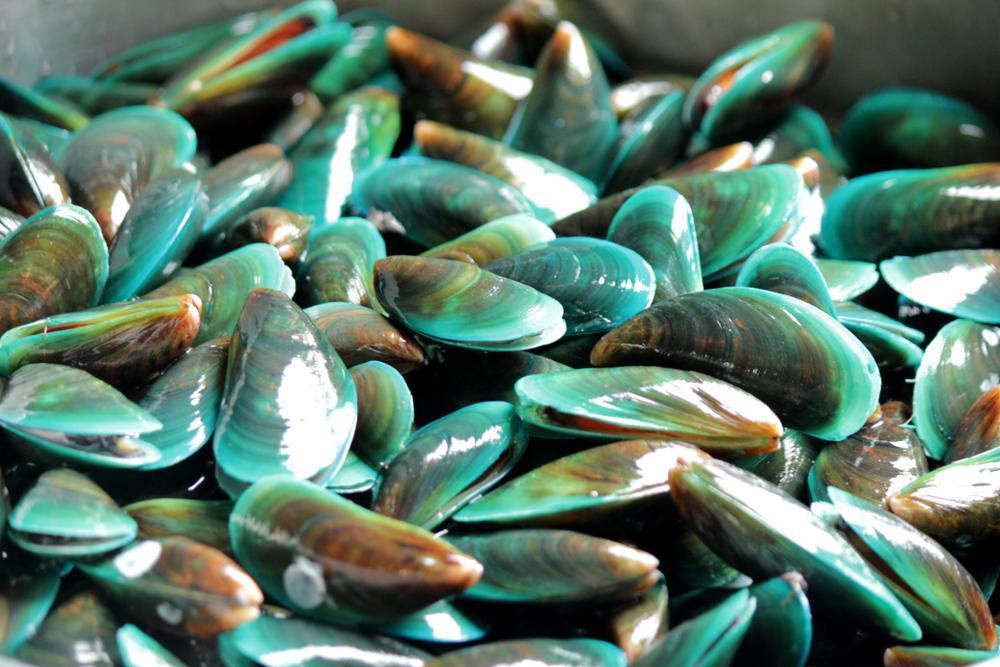
This is one of the questions we are asked all the time here at My Pet Nutritionist. What foods can support joint health in my dog? Well, we thought we’d give you a rundown of 5 of our favourite. 1) Green Lipped Mussels GLM’s contains around 90 different fatty acids, glycosaminoglycans (chondroitin sulphate), glutamine (a glycosaminoglycan precursor), vitamins C and E and minerals including zinc, copper and selenium. The synergy of these nutritional components work in perfect harmony to support joint and mobility issues in pet. When looking at joint health you need to consider all mechanisms responsible for the disease and then how to address them. Inflammation It’s generally the inflammation and rubbing of joints that create pain and stiffness. The fatty acids in green lipped mussels with EPA (eicosapentaenoic acid) and DHA (docosahexanoic acid) are the most abundant. These are the important fatty acids that are known to reduce inflammation. GLM’s are natural COX inhibitors just like NSAIDS (non-steroidal anti-inflammatory drugs), so they can obtain the same pain free results without the negative side effects. They are also natural LOX inhibitors too. Degradation This is the main reason for the breakdown of the joint matrix. The joint matrix and connective tissue need to be supported by components that replicate the structure of joints, naturally. GLMs are rich in glycosaminoglycans and glycosaminoglycan precursors, known as GAGs. An important GAG found in GLMs is chondroitin sulfate a well-documented structural ingredient for joint support. Oxidation Free radical damage can create more degradation and inflammation so we need to support this with natural antioxidants found in GLM. Vitamin C and E are both abundant in GLM’s and essential for joint care. Joint Lubrication Synovial fluid within the joint matrix is incredibly important to support cushioning and movement. Alterations in synovial fluid (SF) lipid composition have been linked to both osteoarthritis (OA) and rheumatoid arthritis (RA). GLMs provide polysulfated glycosaminoglycans (PSGAGs), the building blocks for cartilage and joint fluid. 2) Turmeric Turmeric, Chew-Meric, Tum-eric, no matter how you say it (and yes, everyone seems to say it differently), it packs a whole lot of punch (good punch!) Turmeric is a flowering plant, but it’s the root we’re most interested in. Part of the ginger family, it looks very similar, but it’s the smell that helps you differentiate. When boiled, dried and ground, it’s the spice that is found in many Asian dishes. It has a warm, bitter, pepper like taste with a mustard-like aroma. It’s that spice that makes your hands yellow when you use it! And your worktops, sinks and utensils! It’s known as curcuma longer in the ginger family, and it’s the active compound curcumin that brings the benefits we speak of. It’s often named cure-cumin for the promise it brings. Turmeric is frequently used in humans, to help with a range of diseases and conditions including skin, pulmonary, aches, pains, sprains, liver issues and cancer. Curcumin specifically is argued to be anti-inflammatory, antioxidant, anti-microbial, anti-tumour and also helpful in wound healing. Used in Ayurvedic and Chinese Medicine for centuries, it is now finding a place in Western Medicine. Many joint issues feature chronic inflammation, and in supporting our dogs, we aim to reduce pain and inflammation. So here comes turmeric with its anti-inflammatory properties! Several studies have shown that when supplemented with turmeric, arthritic dogs show a marked improvement in their daily life activity without any side effects! Findings Here It seems to be down to turmeric’s ability to regulate cytokines. 3) Ginger A University of Miami study concluded that ginger extract could be a substitute to nonsteroidal anti-inflammatory drugs (NSAIDs). The study compared the effects of a highly concentrated ginger extract to placebo in 247 patients with osteoarthritis (OA) of the knee. The ginger reduced pain and stiffness in knee joints by 40 percent over the placebo. Research shows that ginger affects certain inflammatory processes at a cellular level, and as we know, many pathologies involving the joints have inflammation as the key player. There are more than 1300 types of ginger plant, and they contain a wide range of nutrients, including: vitamin C vitamin B6 the minerals magnesium, potassium, and copper gingerols, shogaols, paradols, and other phytonutrients and polyphenols Gingerol, shogaol, and paradols all have antioxidant properties, and gingerol and paradols are also anti-inflammatory. Antioxidants help the body get rid of free radicals, which can lead to cell damage and inflammation. Does My Dog Need Antioxidants 4) Oily Fish Joint degradation is characterised by inadequate production of compounds necessary to its structure, along with reduced collagen synthesis. This can be a result of physical stress, trauma, autoimmunity, or aging. Here, inflammation is upregulated, creating further breakdown. It results in weak, damaged, or inflamed tissue with restricted or painful movement. Essential fatty acids are well known to help modulate inflammatory responses found in cases of joint degradation. During the inflammatory response, certain enzymes catalyse the production of compounds which cause pain, redness, and heat. It has been discovered that omega-3 fatty acids inhibit these enzymes that result in this response. Great sources of omega-3 fatty acids include all those oily fish like sardines, salmon, and mackerel. Some plant based oils also contain omega 3 too, hemp seed oil has a great omega 6:3 ratio! 5) Bone Broth Bone broth is a liquid containing brewed bones and connective tissues. Bones themselves are rich in vitamins and nutrients, including calcium, magnesium, and phosphorous. In addition, brewing connective tissue into bone broth provides the body with natural compounds from the cartilage. You will also find collagen and cooking collagen turns it to gelatin, which provides the body with amino acids, which are the building blocks of proteins. It is not possible to say how much of any nutrient will be in a particular batch of bone broth, since this largely depends on the type and quantity of the bones and tissues that went into it, but a good rotation of bones and tissues will provide a range of nutrients. A 2017
There’s a difference between nutrients and foods – nutrients are what you find in foods. Whilst it can be a somewhat reductionist approach to health, if we know the role of certain nutrients, it can help us understand why we need to include them in our pet’s diet. In this blog, we thought we’d look at some of the most important nutrients for bone and joint health in pets. Bone is a light, yet strong connective tissue consisting of around 30% collagen and other matrix proteins with around 70% minerals. These minerals include calcium and phosphorus, but magnesium, sodium and potassium are also present in conjugated form. Bones come together to form joints. The type of joint formed determines the degree and direction of motion. For example, joints with a ball and socket formation allow for a rotation whilst hinge joints only allow for bending and straightening. In a joint, the ends of the bones are covered in cartilage, which helps reduce friction as joints move. With age, this cartilage can degrade. Tendons connect muscle to bone and are made up mostly of collagen. Ligaments surround joints and help to stabilise them. They also connect bone to bone. Bone Health Bone starts as a cartilage model which gets slowly replaced. Osteoblasts are the cells that form new bone; think of it as a blast that spreads, and osteoblasts spread to form new bone. Osteoblasts secrete osteoids which are simply unmineralized bone tissue. Soon after the osteoid is laid down, inorganic salts (calcium and phosphorus) are deposited which forms the hardened material that we know as bone. Inappropriate levels of calcium and phosphorus during growth therefore understandably contribute to bone deformities and skeletal disorders. Calcium Calcium is one of the most abundant minerals in the body. An optimal calcium intake is necessary for bone health at all stages of life. Dietary requirements for calcium are determined by the need for bone development and bone maintenance, which vary throughout life, being higher during puppyhood, adolescence, during pregnancy and lactation, and in the aging. When imbalanced levels of calcium are present, it can result in abnormal skeletal formation and/or function. Causes of Calcium Imbalance Vitamin D imbalance Kidney disease Liver disease Thyroid or parathyroid gland issues Diets rich in phytate and/or oxalate Primary hyperparathyroidism Cancer Certain medications Glucocorticoids promote calcium depletion High sodium diets – when sodium leaves the body it takes calcium along with it Sources of Calcium Raw meaty bones Sardines with bones Salmon Kale (cooked) Chia Seeds Bok Choi Egg Broccoli Liver Vitamin D Vitamin D is actually a hormone that promotes calcium absorption. In human health, you will have heard it referenced as the sunshine vitamin as it is produced in the skin in response to sunlight (UV) exposure. In studies of hip fractures in humans, there appears to be a seasonal variation; more occur during winter months and fracture patients often have low vitamin D status. When supplemented with Vitamin D and calcium, incidences of fractures often reduce. Findings Here https://www.ncbi.nlm.nih.gov/pmc/articles/PMC1240026/ Vitamin D plays an indirect role in bone health by managing calcium levels in the body. It controls absorption of calcium in the intestine and the amount of calcium excreted by the kidneys. If Vitamin D levels are low, then the intestines struggle to absorb calcium. Calcium is key to bone mineralisation (hardening), without calcium, bones are unable to form correctly. Not only that, but due to the lack of circulating calcium, the body mobilises it from the bones by way of increased parathyroid hormone. This not only weakens the bones, but it also creates a new issue, namely secondary hyperparathyroidism. Vitamin D deficiency include symptoms like: Simultaneous deficiency/imbalance in calcium/phosphorus, Rickets (soft and weak bones in young dogs), Osteomalacia (soft and weak bones in adult dogs), Osteoporosis (weak bones leading to fractures), Neurological abnormalities, Hypocalcemia (low calcium levels), Elevated parathyroid hormone (symptoms include bone pain, depression, kidney stones, hypertension, cardiac arrhythmias, and kidney failure), Posterior paralysis, Ataxia (neurological issues including gait abnormality, difficulty walking, tremors), Quadriparesis (weakness in all four limbs). Food Sources of Vitamin D Flesh of fatty fish (salmon, tuna and mackerel) Fish Liver Oils Beef Liver Egg Yolks Joint Health Other than the skeleton, which provides a rigid structural framework for the body, there are other connective tissues that provide support. Where a degree of flexibility is required, cartilage is a rubberlike tissue that offers semi-flexible support for structures. The other function of cartilage is to prevent friction and enable smooth movement around joints. Cartilage is formed by chondrocytes which mainly consist of collagen and proteoglycans. Ligaments are made from tough, fibrous, dense connective tissue. They are made up of collagen, elastin, proteoglycans and a range of minerals including copper, manganese and calcium. Key to proteoglycan structure are the GAG’s chondroitin and dermatan sulphate. Tendons are very similar in structure and function as connectors that join muscle to bone. They are capable of carrying high tensile or compressive forces, facilitating movement around a joint. They have proportionally more collagen and less proteoglycan content as a result of the need for an even tougher structure. Joint degradation is characterised by inadequate GAG, proteoglycan and collagen synthesis to renew tissue in the face of degradation caused by physical stress, trauma, autoimmunity or ageing. Vitamin C Vitamin C is a water-soluble vitamin, antioxidant, and essential co-factor for collagen biosynthesis, carnitine and catecholamine metabolism, and dietary iron absorption. Whilst humans are unable to synthesize it themselves, dogs seemingly can in adequate amounts. Vitamin C is an essential for two enzymes required in collagen synthesis, so sufficient amounts are necessary for optimal joint (and bone) health in the canine. Sources of Vitamin C: Peppers Carrots Pumpkin Sweet Potatoes Seaweed Blueberries Glucosamine Glucosamine is a natural sugar that exists in the fluid around the joints, as well as in animal bones, bone marrow, shellfish, and fungi. The body uses glucosamine to build and repair cartilage. With age, cartilage can become less flexible and start to break down. This