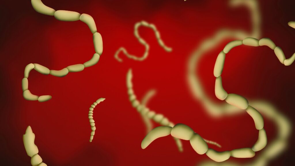
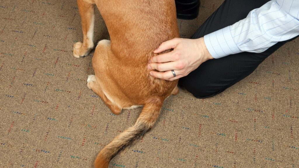
Internal parasitic burdens can be quite worrisome for pet parents – they can affect both our pets, and us humans too, so they’re certainly something to be one step ahead of! Here at My pet Nutritionist, we aim to put your mind at ease, and to help you fully understand the ins and outs of parasites, giving you lots of options to help prevent and control infestations. You can read part 1 here! Welcome to part 2 – Internal Parasites When looking at internal parasites, there are various parts of the body targeted, which we need to look at, including the intestines, (where you can find nematodes, cestodes and protozoa), the lungs, and the heart. Some of these parasites can be life threatening, so it’s important to understand symptoms, prevention, and treatment options. Intestinal Parasites Intestinal parasites are usually referred to by pet owners, as ‘worms’. While some are worms, not all intestinal parasites are worms, nor are all worms equal! Nematodes differ from Cestodes, which differ from Protozoa, so we will delve deeper into each type here in this blog post. Nematodes Nematodes are from the phyla Nematoda, and are your typical worm – they usually look similar to your garden earth worms (though are not actually related!); long and thin, with a squashable texture, and tapered ends. This, of course, is a generalisation, and there are various physical and mechanical differences between the different parasitic nematodes. Here’s the strange thing about nematodes – they can be harmless, and actually beneficial, or they can be parasitic – it’s a very broad phyla! Beneficial nematodes are often used as a means of environmental flea, tick and ant control. These are sold online or from some environmental/gardening shops, and are mixed with water before being sprayed onto the affected area. The microscopic nematodes, now sprayed all over the area, target, and eat the larvae of their target species, which controls pest population! Anyway, getting back to the parasitic kind! Let’s take a look at the intestinal nematodes pet owners may face during their time with pets. Roundworms (Toxocara canis and Toxicaris leonina) Roundworms, also known as Ascarid worms, are very easy to identify in their matured form. If a pet has a burden of these worms, which have matured from their larval stage, they will be very obviously present in the animal’s faeces, or vomit. They are white in colour, usually long (between 4 and 6 inches), thin, and curly – they are often expelled from the body in spirals. The main method of transmission of roundworm to our pets, is through coming into contact with contaminated faeces. Those carrying roundworm, shed microscopic eggs into their faeces, leaving others to come into contact with it. Some insects and other animals, including cockroaches, earthworms, and birds can also carry roundworm eggs, which if eaten by your pet, can pass onto, and mature inside your pet. Puppies can also be born with roundworms, and shed live eggs in their faeces. A question owners often ask, is if they can catch roundworms from their pets – and the answer is yes! Accidental oral contact with your pets faeces can pass roundworms on to you, if your pet is carrying roundworm eggs. Symptoms you may see if your dog has a mature roundworm burden include: Diarrhoea Vomiting Changes to skin and coat Weight loss Bloated appearance to the stomach Visible worms in faeces and/or vomit If your dog has recently contracted roundworm, and it is still in it’s larval stage, it’s unlikely there will be many symptoms, apart from perhaps some loose stools. Findings Here Hookworms (Uncinaria stenocephala and Ancylostoma caninum) The next intestinal nematodes we will look at are Hookworms. As per their name, matured hookworms look like very small, but not microscopic, hooks. They are very thin, no longer than 2cm long, and are white in colour. They can be seen in faeces when they’re matured, but can be confused with undigested food particles, or even small pieces of string. In order to stay inside the intestine, where they are fed and housed in those infected, they have small mouthparts which latch onto the intestine walls where they feed directly from blood vessels surrounding the intestine. There are a few methods of transmission for hookworms in pets – pets can contract them through their mother’s milk as puppies or via the placenta before birth, and they can be orally ingested, or even be contracted through the skin. Hookworm larvae can lay dormant, and reactivate during pregnancy too, so it’s important to run regular faecal samples during pregnancy. Can hookworms transmit to humans? Yes, and no. Larvae can be transmitted in unsanitary conditions, but are rarely transmitted as adults. There is one exception to this however! This may make you shudder, but matured Hookworms can work their way into the human body through the skin, most commonly through bare feet in unsanitary conditions where pets have shed larvae in their faeces. While Hookworm infestations aren’t generally life threatening, they can cause anaemia if left untreated – this is more common in young animals than in adults though. Some symptoms you may notice if your pet has Hookworms include: ‘downward dog’ position in dogs (showing gastrointestinal discomfort) Diarrhoea Vomiting Bloodied stools (due to blood loss when Hookworms detach from the gut lining, as they inject anticoagulants into the localised feeding area) Changes to skin and coat Anaemia, especially in young animals Physical development impairments in young pets Small string like worms visible in faeces Coughing in severe cases If your dog has Hookworms in their larval stage, you may see diarrhoea, but may not see other symptoms, as eggs are microscopic. Findings Here Findings Here Whipworms (Trichuris vulpis) Whipworms, the third nematode sometimes found in our pets, get their name from their shape, much like the other nematodes discussed in this article. If you think of a whip, carried by movie heroes like Indiana Jones, and reduce it’s size to 0.5-5cm – you’ll have
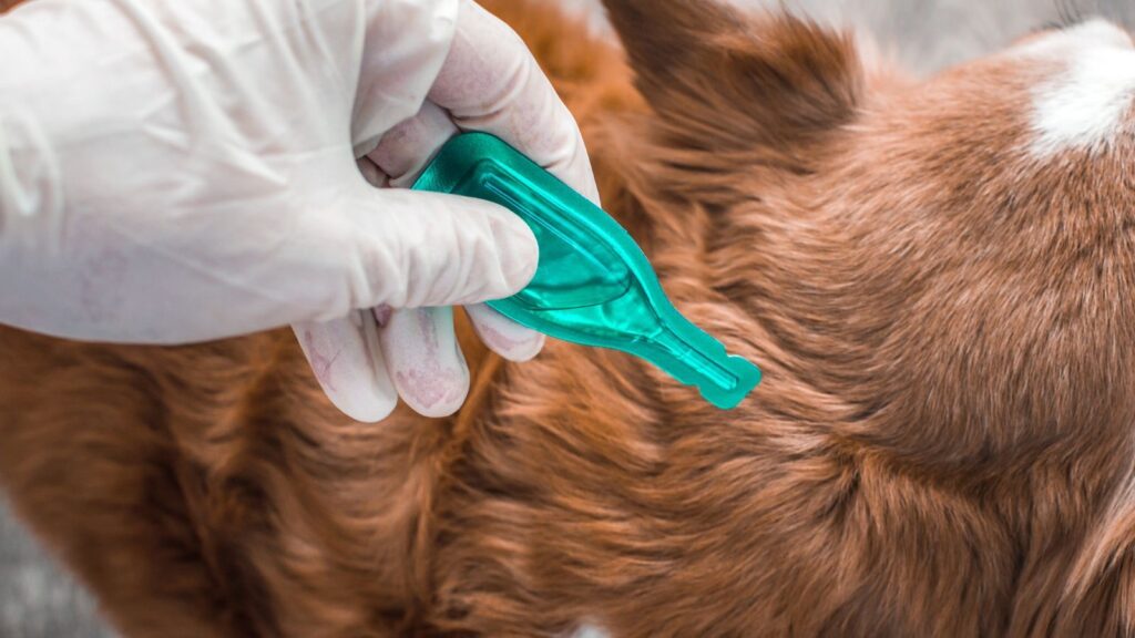
Parasites are always a worry with our pets – let’s face it, our pets touch all sorts on the ground during their walks, sniffing sessions, and play time! It’s inevitable that they come into contact with faeces, urine, slugs, snails and so much more, giving many opportunities for parasites to be picked up, both internally and externally. Here at My Pet Nutritionist we want to help you understand more about parasites, how we can test for them, and how we can help the body rid of them. This is a bumper two part blog, providing you with all the need-to-know information regarding parasites! You can read Part 2 here! Welcome to part 1 – External Parasites Fleas Fleas can be identified as tiny black insects, moving in a hopping form. They jump and crawl around your dog, using a long feeding tube to suck blood from under your dog’s skin. they are very much visible to the naked eye, and another sign of fleas being present, is the dirt they leave behind – this can be identified as black matter in patches across the skin of the dog, at the base of hair follicles. Fleas can be picked up out on walks, or from communal dog establishments when in contact with another dog with fleas, or wildlife with fleas. There are different types of fleas, which target different animal species, some live happily on dogs and cats too, but others can only survive on their intended species, for example hedgehog fleas can only survive on hedgehogs. Cat fleas and dog fleas are the ones most commonly seen in pet households. They can also bite their humans too! If flea infestations are left for too long, the pet can become anaemic (particularly common in puppies), and also suffer with hair loss. Later in this blog post, we will discuss the dangers of using isoxazolines (chewable pest treatments) and spot-on treatments for our pets. Sometimes severe infestations do require one of these products to get them under control, as well as professional house exterminators, but we recommend avoiding these products unless it is as a last resort. Naturally, you can help prevent fleas through the use of selected essential oils (take extra caution with cats when using EOs) and neem oil on the pet, and feeding fresh garlic to dogs (though avoid feeding garlic to puppies under 6 months, and any Japanese breed as these are susceptible to allicin poisoning), as well as another herbal flea prevention supplement. There are many natural topical and internal products on the market, specifically for their target species, whether that’s cat or dog, so using these in a layered approach may give you the best chance of staying flea free! If your pet was to get some fleas, rubbing a thick layer of shampoo onto their dry coat, and leaving for 10 minutes before rinsing out using a comb can really help control the infestation. You can also purchase electric flea-zapping combs which can have great results! In severe cases, food grade diatomaceous earth could be sprinkled onto the coat, however this is extremely drying on the skin, and can be lethal if inhaled. You will also need to focus on steam cleaning your soft furnishings, carpets etc, and clean the house regularly, making extra sure to clean any small, potentially moist nooks and crannies, and wicker items as fleas seem to love these areas! Findings Here Findings Here Ticks Ticks get a really bad reputation – and for good reason, especially in some countries outside of the UK. Ticks can be identified as having a bulbous body, with very small head, and 8 legs, protruding from the head area. When they’re attached to your pet’s skin, you will be able to see the large rounded body (the larger the body, the more the tick has fed), with some legs seen very close to the skin. Ticks need to be removed as soon as possible, as they can carry a range of diseases – anything from Lyme disease (which is the most common tick bourne disease in the UK, though isn’t very common I itself) to disease leading to Alfa Gal Syndrome in humans, and paralysis in the host in other countries. They really can be nasty little creatures! To remove a tick, simply grab a tick removal tool and follow the basic instructions. They’re usually fail safe, and easy to use. If by chance, you do get the head stuck in the pet, these usually work their way out in time. To transmit Lymes Disease, ticks have to be attached for 36-48 hours, however some species of ticks carrying more sinister diseases in countries outside the UK can transmit disease quicker, so it’s super important to check your pets twice a day, and after every walk. You can read more about types of ticks, tick removal, and tick prevention in our blog dedicated to ticks here! Mites Mites come in various forms. The most common ones we see, are Mange (of which there are two types), harvest mites, and ear mites. Let’s take a look at some of these. Mange Mange presents as large areas of hair loss and scabbing. In severe cases, dogs can become completely bald. To test for mange, your veterinarian will take a skin scrape – a sample of the skin’s microbiome, which is analysed under a microscope for mite activity. Each type of mite is a different shape, so the type of mite can easily be diagnosed. Sarcoptic mange is often referred to as Scabies. Sarcoptic mange mites tend to live on the skin’s surface, and tend to be contagious. Ivermectin is the treatment often prescribed by veterinarians to battle sarcoptic mange, however there are some potential complication to using this, which we will discuss later in this blog. Some medicated, anti-seborrheic shampoos, are often used too. There are some natural options that may work in helping fight sarcoptic mange mites, including neem oil, turmeric, and some select essential
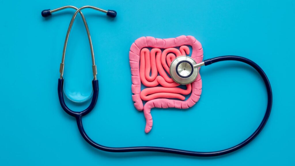
Malabsorption is something we see fairly frequently in dogs here at My Pet Nutritionist. It can be quite scary for pet owners to witness, but there may be some things we can recommend to help these pets. This blog post will explain what malabsorption is, what causes it, and how you can support the body through diet and supplements. What is Malabsorption? Malabsorption is a condition that affects the patient’s ability to efficiently absorb nutrients from food sources. The dog may be consuming a perfectly balanced diet, but still unable to benefit from the nutrition it provides. This is down to underlying problems between the small intestine, and the pancreas; these underlying problems can be a multitude of ailments, so further investigation into potential underlying issues is paramount and working with your veterinarian to discover these, is important. Regular visitors to our blog, may notice we often list breeds who are commonly predisposed to each condition we talk about, however when it comes to malabsorption, there is no specific breed predisposed. This is down to the fact that different breeds will be predisposed to different underlying conditions, and it very much depends on the condition diagnosed, which is causing the malabsorption. Symptoms of Malabsorption There are a variety of symptoms you may notice if your dog has malabsorption. Here are some of them: Pica (consumption of unusual/inedible items) Weight loss, often rapid Lethargy and fatigue Insatiable appetite/extreme hunger Loose stools and gurgling stomach Flactulance Nausea and vomiting Reduced coat condition If these symptoms are present in your dog, please seek veterinary advice as soon as possible as investigations are the important first step in helping your dog with malabsorption. Findings Here Causes of Malabsorption There are many underlying conditions which could lead to malabsorption. We will go through some of these, and give a brief description of each one. You may wish to take this article with you to your veterinary appointment as some of the conditions are not routinely tested for. Inflammatory Bowel Disease (IBD) Inflammatory Bowel Disease is something we talk about, and see a lot here at My Pet Nutritionist. It seems to be very common in lots of dogs who have poor gut health. Those with IBD have inflamed bowels due to the penetration of cytokines and inflammatory cells to the gut and stomach, causing an abnormal immune response. This then has an effect on the lymphatic system, which in severe cases causes Lymphoplasmacytic Gastritis. When the digestive system is inflamed, nutrient absorption becomes very difficult, causing malabsorption. You can read more about severe IBD here. Findings Here Small Intestinal Bacterial Overgrowth (SIBO) We see a lot of patients with SIBO. Those suffering with bacterial overgrowth in the small intestine will often suffer with malabsorption. This major imbalance in the microbiome means that some bad bacteria out-competes the good bacteria required as part of a health microbiome. The bad bacteria, of which there are too many in those with SIBO, often release toxins, and massively interfere with the absorption of nutrients. Findings Here Findings Here Intestinal Blockage Blockages of the intestine are a very common cause of malabsorption. These often go unnoticed, especially if owners don’t realise their dog has swallowed something undigestible, or if the dog is unable to tolerate bone as part of their raw diet. When the bowel is obstructed, food is unable to pass through the intestine, and therefore nutrients are not absorbed from the food, leading to severe weight loss, as part of malabsorption. Blockages causing malabsorption require surgery for removal. Findings Here Exocrine Pancreatic Insufficiency (EPI) This disease is a severe one, and is often not checked for routinely by veterinarians. It may be something to bring up with your vet during your appointment, as though its relatively rare, we are starting to see a few more cases here and there. Your vet may need to do some reading up on it before advising. EPI is occurs due to the pancreas producing less digestive enzymes than needed. Because there is a deficiency in digestive enzymes, foods consumed are not appropriately digested, and therefore nutrients are not absorbed, causing rapid weight loss, large volumes of poor stools, pica, and a variety of other symptoms of malabsorption. This condition is not curable, and dogs diagnosed with it will require regular veterinary check ups, and daily enzyme supplements. Findings Here Findings Here Parasitic Burdens Parasites are another fairly common cause of malabsorption because the parasites compromise the health of the gut, and also syphon nutrients for themselves. Severe infestations of intestinal parasites, or severe cases of giardiasis (where cysts form on the gut following infection from the protozoan parasite Giardia) can lead to malabsorption and severe malnutrition, causing many of the symptoms listed above. It is important to send a faecal sample to a faecal testing laboratory on a regular basis, preferably every 12 weeks, to catch any worm eggs or juvenile worms before large infestations are able to occur. Regular natural worm prevention is also essential for those who tolerate it. You can learn more about natural pest control in our blog here. Findings Here Food Sensitivities Food sensitivities are probably the most common ailments in our customer’s dogs. We deal with a huge number of dogs with food sensitivities, many of which have weight loss as a symptom. Food intolerances are generally coupled with poor gut health, especially cases linked to Leaky Gut Syndrome, whereby the integrity of the gut wall is compromised. When the gut is in bad condition, the microbiome becomes unbalanced, making nutrient absorption tricky. Findings Here Intestinal Growths and Tumours Much like those with an intestinal blockage due to consumption of inedible items, growths and tumours in the intestinal tract are a blockage risk. When these growths or tumours develop and grow, they can create a total blockage of the intestine, which stops nutrients from being absorbed efficiently. While these can be removed by surgery in many cases, some require part of the bowel to removed
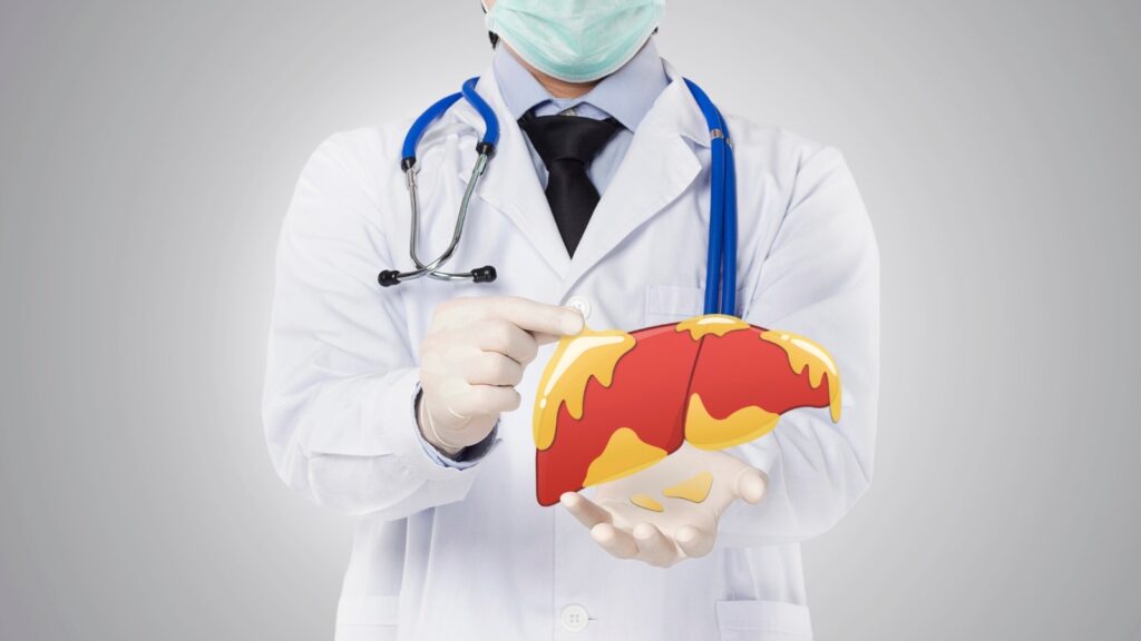
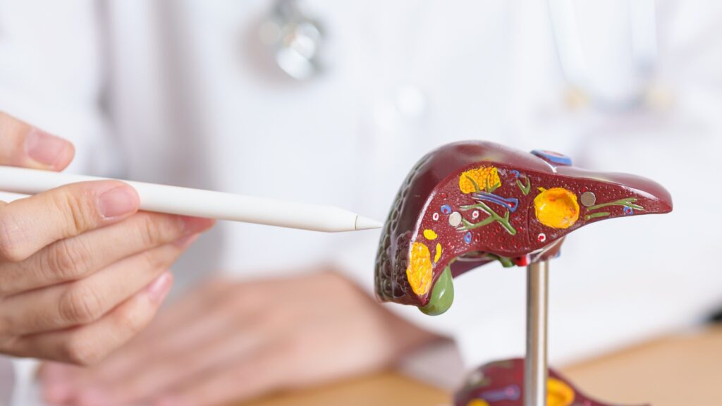
Here at My Pet Nutritionist, Liver Disease is something we help pet parents with quite often. The prospect of living with a dog with liver disease can be a daunting one. But we are here to help put your mind at ease, with this handy blog post packed full of useful hints and tips to supporting your pet’s liver, naturally. The Role of the Liver The liver is an extremely important organ in the body. It plays many roles in the overall health of the pet, including digestion, nutrient absorption, metabolism, detoxification, immunity, and endocrinologically. These roles, all link with one another, much like a large ‘loop’ of bodily functions dependent on the liver. Metabolically, the liver detoxifies fats from the body, and is also involved in the conversion of thyroid hormone 3 to thyroid hormone 4, which we can also look at from an endocrinology point of view. When looking deeper into the liver’s role in detoxification, we see that not only does it detoxify the body of excess fatty acids, but also of drugs, steroidal hormones, environmental toxins, and ammonia (as it helps the formation of urea). In terms of nutrients absorption and storage, the liver is involved in the storage of glycogen, which controls blood sugar levels, and also plays a role in the storage and absorption of Vitamins A, B12, D, E and K, as well as minerals such as iron, magnesium, zinc, and copper. If we look at what the liver produces, we see is produces cholesterol, which is an important precursor to vitamin D synthesis, and production of sex hormones, and it also produces bile, which is used for digestion of food, and also helps keep the gut free from unwanted microbes. Looking at hormone production, the liver produces and secretes four main hormones, or precursors for production of other important hormones. These are: Somatomedin (insulin-like growth factor, essential for regulation of growth of bones and tissues in the body) Angiotensinogen (involved in the regulation of blood pressure and balance of fluid) Thrombopoietin (used by the body to stimulate blood platelet development) Hepcidin (used to block the ability of cells releasing iron, which in turn regulates iron homeostasis in the body) Findings Here Findings Here Findings Here Findings Here Finally, the liver is largely involved in the synthesis of some of the body’s most important proteins, such as: Prothrombin (used in blood clotting) Albumin (plasma protein responsible for controlling the flow of fluids through the capillaries, known as Oncotic Pressure) Globulins (used in immune function) Ceruloplasmin (enables healthy absorption of dietary copper) Lipoproteins (used to transport cholesterol in the body) Findings Here Findings Here Findings Here Findings Here Findings Here What is Liver Disease? Liver disease can be any form of problem with your dog’s liver. Generally speaking, most liver problems in dogs start with an enlarged or swollen liver. Due to the strain on the liver when it is inflamed, liver cells begin to die off, while being replaced by scar tissues – tissues that form after trauma, and can be obstructive if they become too thick. The presence of scar tissues in the liver, cause the structure, texture and appearance of the liver to change. It tends to become firmer – the scientific name for this, is cirrhosis. Cirrhosis is not reversible, and can be a death sentence for the dog, as the liver fails. There can be many triggers and causes of liver disease in pets, including use of some drugs such as selected pest treatments, onset of hepatitis (chronic swelling of the liver), cancer, environmental toxins, infection or sepsis, congenital issues, and even autoimmune disease. Symptoms of liver disease include: Lack of appetite Blood in vomit due to stomach ulceration Jaundice (yellowing of skin, eyes and mucous membranes) Fluid build-up in the abdomen/bloat Excessive urination (polyuria) Excessive water consumption (polydipsia) Diarrhoea Fever Neurologic symptoms Issues with blood clotting There are some changes to diet and lifestyle we can make to help those with liver disease – let’s find out about these! Liver Guard Supporting the Liver Gut Health Gut health may seem like an odd connection to liver disease, however the two can certainly be linked! There’s a group of systems, or axes, which link the gut with the rest of the body, acting as a sort of ‘roundabout’ for the microbiome. You may have heard us mentioning the HPA Axis, the Gut-Brain Axis, and the Gut-Skin Axis; well, the Gut-Liver Axis is another! The main vein involved in the interactions between the gut and the liver, is called the Portal Vein. This is a two way path, through which products derived from the gut are transported directly the liver, and bile, and other liver secretions are transported directly back to the gut to be used in digestion. As the gut microbiome is so incredibly important for prevention, and management of nearly all health conditions, a healthy gut is ever so important. Healthy microbial communities in the gut, make for healthy gut-liver axis homeostasis. Having an unhealthy or unbalanced gut microbiome can lead to exposure to microbes which are pro-inflammatory, causing liver inflammation, and consequently, liver disease. Findings Here To keep the gut healthy, we can tweak the diet, and add supplements – which we will discuss next! Diet As ever, when the body is under inflammatory stress, or has any disease, we need to look at tweaking the diet, to ensure the pet is being fed the most biologically appropriate diet, with the correct nutrition to nurture the body depending on the specific health condition(s) the pet may be facing. A fresh diet would be most suitable, whether that’s raw or cooked. Many dogs suffering with liver problems, may do better on a cooked food instead of raw, but many do just as well on raw! Now, when it comes to different liver conditions, there is no one-recipe-fits-all. Those with liver shunts for example, we would recommend sought advice from one of our team, so we can provide
Here at My Pet Nutritionist, we help pet owners with a host of different health conditions. One we see from time to time, is Spondylosis in dogs. Us humans can get Spondylosis too, but it tends to be more common in our canine companions than it does in their owners. This blog post aims to help you get a better understanding of Spondylosis in dogs, including what it is, how to spot the signs and symptoms, and how you can support the body naturally. What is Spondylosis? Spondylosis, called Spondylosis deformans in the veterinary world, affects the spine; more specifically, the vertebrae – the bones that make up the spine. It’s a degenerative condition, which means it gradually gets worse as the patient ages. Those with spondylosis have bony spurs growing on their vertebrae, known as osteophytes. But why are these growths bad news? The vertebrae protect the spinal chord – probably one of the most important parts of the body as it sends signals between the brain and the rest of the body, and vice versa, and ais also involved in reflexes and coordination. The body spurs growing on the underside of the vertebrae can rub on the spinal cord, causing damage, which then has a knock-on effect to the dog’s sensory capabilities, and ability to move freely. Spondylosis can be widespread, with bony spurs forming along the entire spine, or localised, where they only form in one or two spots. It is most commonly seen in the lower spine, AKA the Lumbar Region, as well as hips and tail. In particularly bad cases, bony spurs can fuse vertebrae together, making mobility tricky. Spondylosis is quite common in aging dogs, especially those who are heavier set, such as giant breeds, or breeds with cobby bodies, and short legs, but it can happen at any age and breed, depending on the cause, which we will look at shortly. The most commonly affected breed is the Boxer, where estimates suggest around 70% of elderly individuals over the age of 9, have been diagnosed with the disease. Findings Here Symptoms of Spondylosis The somewhat strange thing about symptoms when looking at a potential diagnosis of Spondylosis, is that there are very few clinical signs presented, especially not those specific to the condition. Some symptoms you may notice include: Spinal pain: hunching of spine, lameness, unwillingness to walk, difficulty laying down and standing up, general reduction of mobility Behavioural changes: your dog may become more reactive due to pain, and the fear of being hurt by another dog bumping them. Yelping when specific areas are manipulated Lethargy General weakness and stiffness Reduced appetite If your dog is displaying these signs, there could be a number of potential spinal issues, so it’s important to consult your veterinarian to grasp a better understanding of the exact issue your dog is having. Causes of Spondylosis So why does Spondylosis occur? It can be a natural part of the ageing process – just through general wear and tear of an older dog. Another cause of the development of Spondylosis, is trauma to the spine from injury, whether it’s minor trauma, like a bump to the spine, a wobbly landing from a higher surface, or repeated over-use of the spine (often seen in sports dogs who do the same movement repetitively), to major trauma such as having been hit by a car, or having had surgery to correct IVDD. A third potential cause of Spondylosis, is genetic predisposition. There are not many predisposed breeds, but the most commonly seen tend to be Boxers, German Shepherds, and Flat Coat Retrievers. Spondylosis can also form as a secondary disease! Between the vertebrae of a healthy dog, lays soft tissue. This enables the discs to act as shock absorbers, and softens impact, keeping the vertebrae separate. There is a degenerative disc disease, previously mentioned in its abbreviated form, IVDD (Inter-Vertebral Disc Disease), in which the tissue between the vertebrae wears away, leaving bone to rub on bone, prompting the formation of bony spurs to re-stabilise the joint. Findings Here Diagnosing Spondylosis As there is usually a lack of obvious symptoms in cases of Spondylosis, it can be tricky to pinpoint a test for it. If your dog is experiences clear signs of back pain, or even just at the vet for their annual health check where the dog displays possible signs of pain, the vet will use your dog’s medical history records to look for potential missed signs. They will perform any neurological and orthopaedic examinations they feel are necessary, and go from there. Your vet will typically want to rule out any other potential health issues before testing further for Spondylosis. A canine physiotherapist may be able to give a better look into mobility and how your dog’s gait is affected through a video analysis. This may be done through referral; however many allow the owner to book directly in with them to discuss the video, and their findings. Once your veterinarian has ruled out the potential for other, perhaps more common health issues, he or she will book your dog in for an x-ray of the spine, which includes the chest and abdominal cavities, naturally. X-rays tend to give a definitive answer as to whether the dog has Spondylosis or not, due to the visibility of bony spurs on the vertebrae. Less commonly, a vet may recommend an MRI scan – these tend to show potential spinal cord damage as opposed to the bony spurs themselves, which can be an important part of recommending the correct treatment. This procedure is usually carried out on referral with a neurologist. Findings Here Conventional Treatments There is actually very little a veterinarian can do in the event of a dog having Spondylosis, as every individual is different. The treatment plan is heavily based on symptoms the individual’s dog is showing, now a generalised treatment plan, which would be the same for all sufferers. Pain medication is regularly prescribed for those suffering
A question commonly asked by owners, here at My Pet Nutritionist, is ‘how can I boost my dog’s immunity?’, or ‘how do I strengthen my dog’s immune system?’. Well, we’re here to give you some helpful tips on how to strengthen your dog’s immune system – you may be surprised with some of the topics covered, but hopefully you will understand the concepts in this detailed blog post. A healthy immune system is the difference between overall health, and disease, and in many cases, the difference between life or death! We strive to keep our own immune systems strong, and our pets deserve just as much strength in their immune system – they are outside, low to the ground, sniffing public areas barefoot much of the time, after all! The Immune System There are various parts to the immune system as a whole. Generally speaking, when an owner wants to ‘boost the immune system’, it’s the Adaptive (or Acquired) Immune System in question – the immunity gained following seroconversion of vaccinations. Here’s a bit about the Adaptive Immune System, which can be split into two mechanisms: Humoral (antibody mediated) immunity primarily involves B-Lymphocytes. During a humoral immune response, when an antigen is detected, with the help of T Helper Lymphocytes, the B Cells go through a differentiation process, which produces Memory B Cells and Effector B Cells, which are both specific to the B Cell they were differentiated from, and therefore are specifically shaped to combat a specific antigen/pathogen. This is the most common immune response, especially following successful vaccination. Titre Tests pick up these antibodies. Cellular, or cell-mediated immunity involves another type of cell – T-Cells. When T-Cells differentiate, they become T-Killer Cells which attach to and engulf antigens. Allergic responses and autoimmune conditions are part of the cell-mediated adaptive immune system. There are other parts to the overall immune system of a dog, or any other mammal, too. Let’s take a brief look at what these are, and how they differ from the Adaptive Immune System. Active Immunity: this is what is happening when the dog is exposed to a pathogen. The active immune system is the fastest acting system and is the body’s first response to the presence of a pathogen. In the presence of a pathogen, the B-Lymphocytes create and release antibodies. Passive Immunity: this is the immunity passed on to an individual instead of being created by their body. In dogs we call this Maternally Derived Antibodies (MDA). MDA is passed onto the puppies from the mother and is vital to health in the early weeks. It usually wanes between 10 and 16 weeks of age, unless interrupted by vaccination. MDA is passed onto the puppies through the placenta, and through the mothers milk. If a puppy is hand reared, he/she may require extra caution than those who drink mothers milk. Innate (also called non-specific) Immunity: this includes the immunity and defence systems your dog is born with. Barriers like skin, the gastro-intestinal tract, eyelashes etc all help keep pathogens out of the body which is why gut health is so very important – at least 70% of your immune system is in the gut! Defence systems like mucosal layers, saliva, stomach acid etc are also part of the innate immune system. Another immune response included as part of the innate immune system is inflammation – this often acts as a marker to pathogens so they can be destroyed. There are various day-to-day aspects of life, including diet and lifestyle which can affect your dog’s immune capabilities, so let’s dig deeper into these. How Gut Health Affects the Immune System It is becoming more widely known, that 70-80% of the immune system lays in the gut – quite a staggering figure! The all important gut microbiome is naturally perfectly balanced – there are ‘good guys’ and ‘bad guys’ that all make up a healthy gut microbiome, but there’s enough of the ‘good guys’ to keep the ‘bad guys’ under control. When the gut is not healthy, the microbiome is knocked out of balance, and the ‘bad guys’ are able to take over, causing disease within the body. There are various substances, and other triggers such as stress, which cause damage to the gut wall, too. When the gut wall is damaged, the gut microbiome is again, knocked out of balance as it is leaked from the gut (known as Leaky Gut). When the gut is leaking, the immune system is left very vulnerable, so it’s imperative to keep the gut healthy at all times – and if it’s not healthy, focus on getting it healthy! The vast majority of systems in the body are connected to the gut very much like a roundabout. These connections are called axis and there are many, for example, the gut-brain axis, the gut-musculoskeletal axis, the gut-liver axis, and the gut-skin axis. The microbiome communicates with this organ systems by creating messengers and metabolites such as probiotics in order to help support function. This is an emerging area of immunity that science is newly delving into. We will delve into some gut health supplements later on in this blog post. Findings Here Findings Here Gut Guardian How Diet Affects the Immune System The diet plays a large role in gut and immune health. Commercial dry food diets being overly processed do not have the live capacity to nourish the gut microbiome like fresh foods do. Moisture in food is incredibly important for all mammalian species, and dogs are no different. Dry food generally has 6-10% moisture; making it dehydrating to the intestinal tract. We know there are some wondrous foods to feed our dogs to support immunity. Fresh in raw or cooked form is always advisable but there are certain ingredients that we favour as they hold great healing and supportive capabilities. We have a fantastic blog explaining the ingredients we tend to favour, which can be found here. When we look at ingredients, both in kibble and in
Graves Disease is an autoimmune disease involving the endocrine (hormone) system, specifically the thyroid, which we see from time to time here at My Pet Nutritionist. It can be a worrisome diagnosis for any pet owner, and it’s important that owners of pets with Graves Disease understand what it is, and how they can tailor their pet’s diet and lifestyle to keep on top of symptoms; which ultimately extends their pet’s comfort. That’s where we come in, to provide you our Ultimate Guide to Graves Disease in Pets. What is Graves Disease? Graves Disease is the non-clinical, or common, name for autoimmune Hyperthyroidism. You may have heard about Hypothyroidism quite regularly on social media and other platforms where pet owners connect, particularly on dog based platforms; but you may not have seen much discussion on Hyperthyroidism. Graves Disease is rare, but possible, in dogs but tends to be more of a frequent trouble for our feline companions. It can be found in us humans, too! So what exactly is Graves Disease? What happens when your pet has it? If you want the short answer, the thyroid gland in your pet is overactive – it’s working over time! We feel it’s important to understand more about it though, so read on! Graves Disease is an autoimmune condition. Autoimmunity tends to occur due to a damaged immune system, or in individuals with a genetically compromised immune system. The body essentially attacks itself, as it recognises various proteins and other substances in the body, as foreign. The thyroid is a gland which produces various hormones, essential for a healthy life. The hormones produced and secreted by the thyroid are called triiodothyronine (T3), and thyroxine (T4) – these help regulate the body’s metabolic rate. When there is a deficiency in these hormones, important bodily processes slow down, which causes a host of health issues. Now, when these hormones are too abundant, effects on the body can be equally as disruptive. We will discuss symptoms next. Symptoms of Graves Disease There are various symptoms of Graves Disease which you may notice in your cat or dog. Let’s take a look at some of the main symptoms: Weight loss, often rapid Diarrhoea Nausea/vomiting Swelling of the neck and face Increased urination Increased drinking Increased appetite Difficulty swallowing Shortness of breath and increases heart rate Change in pitch of bark Reduced eyesight capabilities Behavioural changes, including anxiety depression, and/or hyperactivity. Causes of Graves Disease The cause of Graves Disease is very much unknown. There is very little research into the causes of Graves Disease. It is often describes as ‘idiopathic’, meaning there’s no known cause. Some studies suggest that one known cause of Graves Disease in dogs (though rare) is as a result of a rapidly spreading cancer, called thyroid carcinoma. In cats, some cases of Graves Disease are secondary to a non-cancerous tumour forming on the thyroid gland called Adenomas. Like in dogs, in rare occasions, malignant tumours known as adenocarcinomas can cause the development of Graves Disease by causing the over production of hormones. It is possible that deficiencies in the diet may contribute to onset of Hyperthyroidism, as well as exposure to chemicals and toxins absorbed by the body, including flea and tick products which are toxins, and end up in the bloodstream, which disrupts the normal functioning of the thyroid. Diagnosing Graves Disease The first step in a diagnosis of Graves Disease, is a physical examination of the neck area of your pet. The purpose of this, is to feel for an enlarged thyroid gland. Secondly, the vet will take a blood sample to test for hormone levels in the blood. If the reading comes back with a value higher than the ideal’ threshold for each hormone, it’s very likely your pet has Graves Disease. If the T3 readings are high, but the T4 readings are ideal, more tests may be required, which would be discussed with the vet. These tests may include further bloodwork, and a urinalysis, which will show potential secondary defects to the heart and kidneys. As the thyroid is involved hormonally with the vast majority of the body’s organs, imperfect bloodwork in relation to other organs, could help show a problem with the thyroid. Conventional Treatments As always, here at My Pet Nutritionist, we like owners to be able to make an informed choice with their dog’s health, and encourage the use of conventional treatments where necessary in life potentially endangering situations, and situations where quality of life becomes the biggest importance. This is of course, alongside as much natural support as possible, and feeding a fresh, therapeutic diet. More on this later – let’s take a look at the conventional treatments your veterinarian may offer. There are various approaches which may be taken. As carcinomas are one of the most common, treatable reasons a dog or cat may have Hyperthyroidism, most are based around treating the cancer. Radioactive Iodine Therapy Radioactive Iodine Therapy is fairly common in practices which offer it as a treatment. The radioactive iodine is injected into the bloodstream, which makes its way to the thyroid gland. The thyroid gland takes the iodine up, causing radiation to be emitted. The emitted radiation then destroys any cancerous tissues around the thyroid, but does not cause any damage further afield. This treatment often results in normal levels of hormone production within mere weeks of the injection, making it extremely effective in curing hyperthyroidism. This treatment option has to be undertaken in a specialist veterinary hospital with licencing to carry and use radioactive substances. The pet must also be kept in the surgery until their levels of radiation are safe for handling. Findings Here Findings Here Medication Some drugs may be offered, to inhibit the production and release of hormones from the thyroid. As the thyroid is overactive, slowing it down is imperative to managing Graves Disease. This method will not cure, but can support the pet in reducing flare ups and secondary health implications. Being the
There are so many aspects of health that we look at here at My Pet Nutritionist. Diet and lifestyle are considered, including vaccination schedules, chemical treatment exposure, the quality of water provided and the cleaning products/household products used in the pet’s environment. However there is another factor often overlooked when it comes to disease – stress! We often say disease is expressed according to how we interface with our environment. The main factors here being nutrition, toxins and stress. Stress can be mental, or physical. Mental stress and physical stress can also be linked. In this blog, we will look at the links between stress, and disease. What is Stress? We all know what stress feels like from time to time (and if you don’t, the rest of us are very jealous!), but how does it look from a biological standpoint? What actually happens in our and our pets’ bodies during stress? Stress responses start off in the part of the brain that deals with emotions; the Amygdala. The Amygdala sends a message to the ‘control centre of the body’; the Hypothalamus, the very centre of the brain. The Hypothalamus then interacts with the autonomic nervous system (the part of the nervous system that controls involuntary bodily functions such as breathing, heartbeat, blood pressure etc) to make adjustments in the body, to cope with the stress. The autonomic nervous system is the main system involved in stress responses, and branches into the sympathetic, and parasympathetic nervous systems. These systems are what give the tell-tale signs of stress in your pets (and yourself!), and what help reduce the effects of the stress response. Let’s take a look at some of the common symptoms of stress, caused by the different systems: The sympathetic nervous system Inhibits saliva production Increases heartrate Dilutes pupils Relaxes urinary bladder Inhibits digestive capabilities (read on to find out more on this!) Dilates bronchia The parasympathetic nervous system Promotes saliva production Decreases heartrate Constricts pupils Constricts urinary bladder Stimulates digestive functions Constricts bronchia Stress isn’t always emotional – it’s important to remember this! Common stressors include: Emotional: fear, mental trauma, anxiety Physical: over-exertion, injury, pain Environmental: allergens, pollutants, radiation and rapid temperature changes Biological: bacteria, viruses, parasitic burdens Chemical: pesticides/herbicides, toxins, heavy metals Consumable: ultra-processed foods You can read more about the stress response here! Findings Here Findings Here Calm Complex The Gut-Brain Axis Gut health plays a role in mental health, which means the health of your pet’s gut, is incredibly important to look at if your pet is frequently stressed. The gut is very much linked to every system in the body, and the nervous system is no exception! Bidirectional (both ways) occurs between the gut and the brain, so having a stressed pet, can be a little like a vicious circle, in that emotional stressors can affect the gut, and poor gut health can affect the pet’s emotions. Located in the peripheral nervous system, the main nerve associated with this bidirectional signalling between the gut and the brain, is the Vagus Nerve. Unlike other cranial nerves, which signal between the head and neck areas, the Vagus Nerve reaches all the way through the body, to connect the brain to the gut. The Vagus Nerve is responsible for various bodily functions, including: Allowing for swallowing and vocalisation in the larynx and pharynx Parasympathetic supply to the heart in the thorax, which reduces the heart rate during stressful situations Regulates smooth muscle contraction in the intestine, to enable normal defecation The Vagus Nerve is essential to link the central nervous system to the enteric nervous system to enable healthy digestion. You can read more about the Gut-Brain Axis here! Findings Here Emotional Stress and its Effects on the Body Having an anxious pet can be heartbreaking for the owner – not to mention hard work (which is very much worth it!). Due to the gut-brain axis, we know that emotional stress, doesn’t just stay within the brain! It can cause disease throughout the body, purely down to the fact the Vagus Nerve is an important part of so many systems in the body. The endocrine (hormone) system, and enteric nervous system are both massively affected by poor brain health, and prolonged periods of emotional stress can lead to a host of hormone-related diseases, and disease within the digestive tract. Of course, not all dogs with endocrine or digestive issues are stressed, nor can it always be put down to stress, but stress responses and brain health are very often overlooked – so here is your reminder to check your pet’s mental wellbeing. Think about things you could improve in their life to give them a calmer, more level mental state if they are typically easily overwhelmed, or provide them with a more stimulating routine or space if your dog’s mental health is poor due to boredom. It works both ways! Read on to find out more on how to keep your pet’s stress levels to a minimum. Findings Here Calm Complex Physical/Environmental Stress and its Effects on the Body When our pets go through some form of physical stress, whether it’s injury, or illness, the pressure on the body’s systems (which are already working harder than normal, in order to fight disease or heal injuries) can once again, cause a vicious circle. The added pressure on the body’s systems, makes for the potential for disease to worsen due to stress. We need to help our pets recover in a timely manner with as little emotional stress as possible, in order to reduce stress. Physical and environmental stress can cause emotional stress, which we know can lead to endocrine and digestive upset. Gut damage can then lead to numerous other diseases because 70-80% of the immune system lays in the gut. Chemical/Consumable Stress and its Effects on the Body Chemicals used on the pet, and around the pet as well as what you feed your pet can cause stress on the body, which leads to disease. This is
Here at My Pet Nutritionist, we see a lot of people worried about their dog’s recent blood test results. The Liver Enzyme reading may be higher than expected for a variety of reasons, and could be partly down to the food being fed to your dog. This blog aims to help you gain a better understanding of the value you may have received, and what may have caused it to be high. A Bit About Liver Enzyme Readings When your dog has a full panel of blood tests, performed by your veterinarian, your dog’s liver function is tested. The results will show on the results sheet as values for ‘ALT’, ‘AST’, ‘ALP’, and ‘GGT’. There is a set range of values which denote normal/healthy liver function which are as follows: ALT: 12-118 U/L AST: 15-66 U/L ALP: 20-200 U/L GGT: 0-25 U/L Each reading is the result for a different liver enzyme – read on to learn about each one. Your vet should help you analyse your dog’s results, and offer feedback as to the health of your dog’s liver. Findings Here Findings Here Findings Here What are the Liver Enzymes? As mentioned, each value relates to the levels of a different enzyme. Enzymes help to speed up chemical reactions in the body, and in the case of the liver enzymes as a whole, they are responsible for the production of bile, blood clotting, digestion, breaking down toxins, and helping the body to fight infection. Let’s take a look at the different enzymes tested for in your dog’s liver. Alanine Transaminase (ALT) ALT is an important catalyst for the process involved in the metabolism of glucose and protein, to for pyruvate (major part of cellular respiration) and glutamate (an important neurotransmitter used for memory, mood regulation and cognitive capabilities). When liver cells are damaged, ALT is released into the bloodstream. High amounts of ALT Leakage, means your dog could have liver disease or trauma. Findings Here Findings Here Aspartate Transaminase (AST) AST plays a major role in Gluconeogenesis of the liver, as well as some other tissues and organs. It is a catalyst for the transfer of an amino acid from aspartate to glutamate. Raised AST values can also be caused by leakage into the bloodstream through disease or trauma to the liver, as well as trauma to the muscles in the body, as AST is also found in the musculoskeletal system in mammals. Findings Here Findings Here Alkaline Phosphatase (ASP) ASP is found throughout the body, in the bloodstream. It is the catalyst for the hydrolysis of phosphate esters, leading to the breakdown of proteins in the body, and it is produced not only by the liver, but in the kidneys, intestines, pancreas and bones; it is however, produced mostly by the liver. Findings Here Findings Here Gamma-Glutamyl Transferase (GGT) GGT aids the transfer of amino acids through the membrane of cells, and is also involved in leukotriene metabolism. It is a prominent marker of liver dysfunction, so elevated GGT readings are best to be further investigated as soon as possible! Findings Here Findings Here Liver Guard What Causes Raised Liver Enzymes? While there are various underlaying health conditions which cause raised liver values, including liver disease, hepatitis, cancer, thyroid disease, various myositis diseases (muscular disease), diet can influence your dog’s liver enzyme values. Too Much Copper When there is too much copper in the diet for the individual dog, the dog may develop copper-storage liver disease, scientifically known as Copper Hepatopathy. Copper is an important nutrient to include in your dog’s diet, as it aids the production of energy, and maintenance of blood vessels, and connective tissues throughout the body, however the liver is unable to process large amounts of it, which leads to a build up in the liver, which then has to store it. This damages the liver, causing elevated liver enzymes due to disease. Findings Here Findings Here Low Copper Too Much Vitamin A While Vitamin A is an important part of a balanced diet, essential for ocular health, healthy development, a strong immune system, and reproductive health, supplementing the diet with extra Vitamin A can cause elevated liver enzymes. When looking at nutrient profiles of your dog’s diet, try to use fresh ingredients (fresh fruit and veg, specifically selected, may be the best addition to your dog’s diet), instead of a supplement. Supplements tend to be very concentrated, which could easily tip your dog over their recommended daily intake of Vitamin A – this is called hepatotoxicity. Findings Here Findings Here Findings Here Too Much Iron Over-supplementing iron can also cause hepatotoxicity, which occurs due to iron-poisoning. Having excess iron intake causes raised liver enzymes due to the damage caused by hepatotoxicity, known as hemochromatosis. Findings Here Findings Here Zinc Deficiency Zinc deficiency often comes hand in hand with excess Vitamin A consumption. Some studies suggest that elevated Vitamin A in the blood could be caused by a lack of zinc, not just by over-consumption. Zinc levels are often skipped during testing for various diseases, but are extremely important to investigate. Findings Here Findings Here Findings Here High Carbohydrate Diets Studies show that dogs fed a high carbohydrate diet are more at risk of developing liver disease, and therefore raised liver enzymes than those fed a low carbohydrate diet. One of the roles of the liver, is to maintain glucose concentrations in order to control the metabolism of carbohydrates. If the liver is strained too hard due to being overloaded by carbohydrates, it is at risk of leaking enzymes into the bloodstream, causing elevated liver enzymes. Findings Here Findings Here Findings Here Ketogenic Diets As much as a high carbohydrate diet may lead to leakage of liver enzymes, a low-to-no carbohydrate diet may also lead to the leakage of liver enzymes, as the liver overproduces them. Some conditions do require a ketogenic diet, such as those suffering from cancer, but if your dog is on a ketogenic diet, ensure your dog’s blood is
Here at My Pet Nutritionist, we deal with a huge amount of dogs with skin complaints. One of the skin complaints we see fairly often is Furunculosis. As a recurring skin infection, there are things we can do to support the body; read on to find out how we can help. What is Furunculosis? Furunculosis is a deeply embedded infection of the skin, or deep rooted inflammation of the skin. it is a bacterial infection of the skin and soft tissues, and emanates from the around, and inside the base of hair follicles, reaching through the dermis, and into the subcutaneous layers of the skin (the layers just beneath the skin’s surface). Findings Here Furunculosis clinically presents as small, often pus-filled boils on, or just under the skin. There are a few types of furunculosis you may have seen mentioned in your time as a dog owner, so let’s take a look at the common ones! Anal Furunculosis What is it? Probably one of the most common types of furunculosis mentioned by dog owners, is Anal Furunculosis. As per it’s name, this is where hair follicles around the anus become infected, and chronically inflamed. Another name for Anal Furunculosis is Perianal Fistula. This is an extremely painful type of furunculosis, as well as being one of the most difficult to treat, and stop from recurring. Findings Here Causes Evidence suggests it’s an immune mediated disease, as there is a strong genetic association with one specific allele, and tests show upregulated cytokine expression causing T-cell infiltration. It also tends to mostly affect middle aged dogs. Findings Here Findings Here A zinc deficiency could contribute to the onset of furunculosis too; zinc deficiencies usually present clinically in the form of a variety of skin issues. Findings Here Genetic predisposition is a common cause of anal furunclulosis, including the following breeds: German Shepherd (most common predisposition) Beagle Labrador Various Bulldog breeds Old English Sheepdog Australian Shepherd Staffordshire Bull Terrier Findings Here Symptoms There are various symptoms of anal furunculosis, but many are similar to general anal gland troubles, and not obvious until the infected boils begin to show. Some signs and symptoms include: Scooting Licking/nibbling around the anus and base of the tail Change in posture Difficulty passing faeces Bloodied stool (bright red) Lethargy Potential weight loss Interdigital Furunculosis What is it? Many of our readers will likely have seen ‘interdigital cysts’ mentioned by other dog owners – these are incredibly common in our domestic dogs. ‘Interdigital cyst’ is the somewhat shorter, almost self-explanatory term for Interdigital Furunculosis. Interdigital means ‘between the digits’ – no, not number in this sense; but the toes. Inflamed and infected boils/cysts appear between the toes, causing pain and annoyance to the dog, due to infection showing outwardly, from the hair follicles in the area. Findings Here Causes Most causes of Interdigital Furunculosis are down to trauma to hair follicles present between the toes. A variety of things can cause this, which we will discuss below, but first, we will take a look at some other potential causes of interdigital cysts. Endocrine diseases such as Cushing’s Disease and hypothyroidism can contribute to problems internally with the hair follicles around the toes. Bacterial and fungal infections in the foot areas, including yeast (for which you can read more about in our blog here), and various strains of Staphylococcus infection, can cause the onset of furunculosis in between the toes. Now on to the trauma related causes! Paw licking is a huge risk factor for hair follicle trauma between the toes. Paw licking can be caused by food sensitivities, anxiety, pain in the area, or even due to compulsive neurological disorders. Foreign bodies like the dreaded grass seed may also cause both paw licking, and generalised inflammation of the toe area, and therefore increase the risk of interdigital furunculosis. The root cause of any paw licking must be found and treated as a matter of importance! The length of a dog’s paw hair can also cause trauma to the hair follicles. When the dog walks on his or her feet, the hair between the toes may rub, which causes skin inflammation, and leaves the hair follicles vulnerable to infection. Those with longer fur between the toes, may be at a much lower risk of hair follicle damage, as the fur helps protect the follicles. Dog breeds at a greater risk of interdigital irritation due to their shorter coat length include: Various Bulldog breeds Basset Hound Staffordshire Bull Terrier Pug Great Dane Boxer Boston Terrier Beagle The shape of the dog’s foot can also have bearing on it’s potential for the onset of Interdigital Furunculosis. Those with wider gaps between their toes leave the skin in between their toes open to irritation by the environment. Those who are overweight are also more likely to have poor paw conformation whereby their toes spread, causing potential for irritation of the interdigital hair follicles. Findings Here Symptoms Early signs of Interdigital Furunculosis are a little easier to spot than Anal Furunculosis. Generally, the area will become red to start with, and slight swelling may become apparent. In cases where a foreign body is present, oozing of the area may occur. This needs veterinary treatment right away. The dog will usually lick, nibble and or favour the paw(s) affected, which is a very strong sign of a paw ailment. Once fully established, the Interdigital Furunculosis will present as a large swelling between the toes, usually warm or hot to touch, and very red in colour. It can be a shiny texture on it’s surface too, due to the swelling of a specific hair follicle, and lack of fur on it. Findings Here General Furunculosis After Grooming What is it? While there’s generally no single reason for the onset of both Anal and Interdigital Furunculosis, generalized post-grooming Furunculosis has a clear trigger, and usually crops up 24-48 hours after a dog has been groomed. It can establish in any part of the body, particularly in
Here at My Pet Nutritionist, we see many dogs with severe gastritis. There are so many types of gastritis, and Lymphoplasmacytic, or Lymphocytic-plasmacytic, Gastritis is one of the common types of gastritis we see. It’s quite a mouthful of a word, so hopefully this blog should simplify it, and give our readers a deeper understanding of the condition, and how we can help through food, lifestyle changes, and supplementation. What Is Lymphoplasmacytic Gastritis? Don’t let the length of the word scare you – you may not have heard of Lymphoplasmacytic Gastritis before, but it’s likely you have heard of IBD; Inflammatory Bowel Disease. Lymphoplasmacytic Gastritis is a chronic form of IBD, whereby inflammatory cells and cytokines (the substance which stimulates inflammation of cells) penetrate the stomach and intestinal lining. As you can imagine, when inflammatory cells enter the digestive system, it causes havoc! The inflammatory cells invade the stomach and intestinal lining due to having been subject to an abnormal immune response. There is a huge link to the lymphatic system in the gut too; which means a knock on effect to the rest of the body is very likely. This is where the name ‘Lymphoplasmacytic’ comes from – the link with the lymphatic system. Lymphoplasmacytic Gastritis happens most commonly in older dogs, but has been known in dogs as young as 8 months old. There are a number of conditions which have very similar symptoms to Lymphoplasmacytic Gastritis, including Giardia, a protozoan parasite we see regularly, Salmonella poisoning, Pancreatitis, and Small Intestinal Bacterial Overgrowth (SIBO) which we also see frequently. A more severe health condition with similar symptoms is Lymphocytic Gastritis-Like T Cell Lymphoma. These similarities make diagnosis a little tricky. Findings Here Findings Here Findings Here Symptoms of Lymphoplasmacytic Gastritis This leads us nicely on to spotting the signs and symptoms of Lymphoplasmacytic Gastritis. There are a variety of symptoms of Lymphoplasmacytic Gastritis, which are as follows: Infrequent flares to start, gradually becoming more frequent as time goes on Chronic diarrhoea Nausea and Vomiting Bloody vomit Loss of appetite Weight loss Abdominal swelling Dark, bloody stool Coughing up blood Lethargy Some dogs may struggle to breathe Diagnosing Lymphoplasmacytic Gastritis As with any symptomatic patient, it’s incredibly important to visit your veterinary surgery, and have a consultation with your veterinarian in order to properly diagnose the condition and work out your next plan of action. Your vet will take a sample of blood in order to run a full blood panel to look for potential issues with your dog’s blood values. The blood values are commonly within normal ranges for dogs suffering with this condition, but some are slightly anaemic, or have lower than normal levels of proteins in the blood. A test for pancreatic function may be carried out to rule out the possibility of pancreatitis, and Vitamin B12 and Folate levels may be tested; this checks the intestines ability to absorb efficiently. You can read more on folate and B12 levels in our blog here! The final, and possibly most helpful step in diagnosis of Lymphoplasmacytic Gastritis, is scanning. While standard x-rays tend to look ‘normal’, ultrasounds will show inflammation in the bowel area, and are often the key to diagnosis. Some vets may carry out a Barium radiograph, which is more useful than a standard x-ray, but not preferable to an ultrasound. For definite confirmation of Lymphoplasmacytic Gastritis, a biopsy will be taken. Findings Here What Causes Lymphoplasmacytic Gastritis? There are various causes of Lymphoplasmacytic Gastritis. Let’s take a look at them! Breed predisposition: some breeds are genetically predisposed to the condition. These include the cocker spaniel, basenji, shar pei, German shepherd, Yorkshire terrier and wheaton terrier. Food sensitivities: always a big topic here at MPN, food intolerances and allergies can be a huge factor for the development of Lymphoplasmacytic gastritis. SIBO (Small Intestinal Bacterial Overgrowth): bacterial overgrowth damages the gut, which in turn leads to inflammation Parasites: parasitic infection can lead to major inflammation in the gut and bowel. Bacterial infection: having a bacterial infection causes an imbalance in the gut microbiome, which has a huge knock on effect within the body. Inflammation is a major issue caused by this. Findings Here Findings Here Conventional Treatments Of course, there will be pharmaceutical approaches your vet may offer. Once independently researching these, you may wish to proceed, but you may wish to support the body naturally. We cannot sway your opinion either way, so it’s important to listen to your veterinarian, and fully research recommendations, particularly longer term recommendations. It’s likely your vet will prescribe a binding medication, which helps bind the contents of the bowel – these pastes usually contain a beneficial clay called Kaolin, and a probiotic to help repopulate the gut. Anti-nausea medication may be essential in the initial recovery of your dog, to enable the dog to stop vomiting, and hopefully encourage eating. As your dog will likely be dehydrated due to the chronic diarrhoea and vomiting they will likely suffer, it will be important to rehydrate through intravenous fluids. IV fluids will be carried out at the veterinary practise. To help the initial recovery, your vet may also prescribe antibiotics in case of infection, steroids to help reduce inflammation in an effective and timely manner, and diuretic medication to reduce the amount of excess fluids in the body. After the first round of medication, your dog will be reassessed, and further treatment decided upon, if necessary. Your vet may offer an antiparasitic medication at this point, however we would advice a full faecal panel first, as there’s no point treating something that is not there! Your vet may discuss dietary changes to help reduce the risk of reoccurrence, and this is where we can help! Findings Here Findings Here Supporting the Body Naturally So how can we support the body naturally, to keep on top of Lymphoplasmacytic Gastritis? Diet Diet plays a large role in supporting the body. As always, fresh food is most suitable for those with Lymphoplasmacytic Gastritis.
Yeast is a problem we see very often here at My Pet Nutritionist. Owners often feel they have run out of options with regards to treating the yeast, so this guide has been made with the goal of helping you overcome yeast; you may just see something missing from your routine which could be the missing piece for success! What Is Yeast? Yeast, as we commonly call it, comes in two forms – Candida; a fungal pathogen, and Malassezia; a skin based hydrophobic yeast. Candida makes up a small portion of a healthy gut microbiome, and is naturally found in the gut, and genital tract. While candida is a very normal part of a healthy gut, if the gut is not kept in tip top condition, it can cause problems. In the healthy gut microbiome, there are good bacteria, and there are bad bacteria. The good bacteria keep the levels of bad bacteria in check, which means the bad bacteria aren’t able to wreak havoc! When the gut is impaired the opportunistic fungal pathogen, candida will take hold, and grow. Now we have too much of the ‘bad guys’ and not enough of the good bacteria to fight it off, and so candida takes over! Once candida has taken over, and the gut is not healed, we start to see the typical yeast symptoms in our pets. Malassezia often happens as a result of environmental allergies, as the skin barrier is damaged, allowing for the formation of yeast on the skin. Much like inside the gut, the skin has it’s own microbiome, in which the bad pathogens are kept in check by the good. When the good pathogens diminish, the bad ones are able to take over! This is something we see in a vast amount of customers who come to us with a dog with environmental allergies. Symptoms include: Rusty colour, mainly between toes and paw pads, ears, under the tail, and around the groin and armpit areas. It can occur in other places too, but these moister areas are prime location for it! Repetitive paw licking Licking under the tail area Ear scratching and head shaking Excessively rubbing face and body on walls and furniture Hair loss An unmistakable odour; much like a damp, cheesy smell, or popcorn-like smell Sometimes discharge is apparent in yeast cases More Information on yeast can be found here. Let’s take a look at how we can help our dogs battle Candida and Malassezia! Battling Yeast When dealing with yeast, there are many contributing lifestyle and nutrition factors, which may need to be tweaked a little, or added to combat the pathogens, and repair both the gut barrier, and the skin barrier. Diet As always, here at My Pet Nutritionist, we are very fresh food forward. Feeding a dog with yeast is no different – one of the main steps to battling yeast, is cutting out dry food as much as possible. A fresh, balanced diet would be your best option whether it’s raw, or homecooked using one of our balanced recipes. Why does diet matter for those suffering from yeast? Let’s take a look at the composition and manufacturing process of dry foods, first and foremost. Kibble is high in carbohydrates: generally speaking, most kibbles are between a whopping 30 and 70% carbohydrates! Carbohydrates are sugars – a fantastic food source for yeast! The canine body struggles to digest carbohydrates often found dry foods, which causes gut inflammation. Kibble is ultra processed: excessive processing causes the food to become inflammatory. When the gut becomes inflamed, damage occurs, which leaves the microbiome vulnerable, and causes imbalances as the bad microbes out compete the good microbes. Advanced Glycation End Products (AGEs) are produced during high heat manufacturing and extrusion of dry foods. AGEs not only speed up the ageing process, but they have many other health disadvantages too, one being that they expand the life of yeast. We need to cut out products which produce AGEs when we are battling yeast! Findings Here Why can fresh feeding help combat yeast? You can tweak the diet to ensure there’s no starchy carbohydrates. Less ‘food’ for the yeast! It’s much easier on the gut, as it’s highly digestible! This means less gut damage and less inflammation. There’s no high temperatures or vigorous processing involved, meaning no AGEs form! What do we need to include in a fresh diet for yeast sufferers? Our recipes for cooked foods are already balanced for you! However if you are raw feeding, you will want to feed 80% muscle meat, 10% bone, 10% secreting offal (half liver, half other secreting offal, preferably!) with the addition of omega 3 sources like raw eggs, fish oils and oily fish, and algal oils. Some vegetables may be suitable to be added to the diet of a yeasty beastie, but we need to be very selective, and very careful which ones we pick! High fibre, low starch veggies are ideal, if tolerated – it’s wise to start off by giving a miniscule amount of plant matter, and gradually building up to 10% plant matter. Some options are broccoli, kale, and cabbage. Try to avoid starchy carbohydrate plants such as sweet potato, carrot and squash. Supplements There are so many supplements on the market these days – it can feel quite daunting selecting the most suitable ones for your pet. Some are better than others when it comes to battling yeast. Gut healing: gut healing supplements are very important – in order to regain balance in the gut microbiome, we want the gut to be in a good, healthy, and strong state. Mucilage herbs are our friend here! Slippery Elm, Marshmallow Root and Deglycyrrhizinated Liquorice are all great for this purpose! Gut Guardian Probiotics: probiotics are essential for yeast sufferers, as they help to rebalance the gut microbiome, by out competing the bad bacteria, which in turn will aid the recovery of the skin barrier. Once there are sufficient levels of good bacteria, the ratio