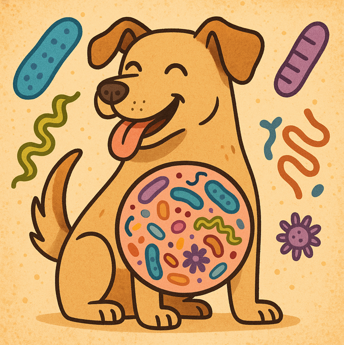
Here at My Pet Nutritionist, we often have queries regarding health and it’s relation to behaviour. The vast majority of reputable behavioural professionals will refer you to a vet and or nutrition specialist such as us in the event of behavioural issues. With strong links between nutrition, health and behaviour, your pet’s diet and wellness regime is important to take into consideration when battling a behavioural issue, or preventing them. Read on to find out how health and nutrition affects behaviour. Microbiome The gut microbiome consists of various good and bad bacteria, viruses, fungi and parasites. These pathogens can be split into the ‘good guys’ (good bacteria), and ‘bad guys’ (the rest!), however in a healthy gut microbiome, the good guys outweigh the bad guys, which keeps potential disease at bay. When the balance of microorganisms in the gut is off, the bad guys take over and disease occurs, as well as behavioural problems. The gut is directly linked to behaviour via the gut-brain axis. This is a pathway, which is one of a series of pathways/axis linking parts of the body to the gut, which connects the gut and the brain. If gut health is poor, brain health is poor. A dog with behavioural issues such as anxiety can display gut issues such as diarrhoea. These are the common gut health problems which contribute to behavioural difficulties: SIBO (Small Intestinal Bacterial Overgrowth) happens when the small intestine has too much bad bacteria from the large intestine, which overtakes the good bacteria. This poor balance in the microbiome plays a part in disruption of the endocrine system and can impact the production of neurotransmitters, often leading to anxiety, aggression and the inability to settle. Dysbiosis is different to SIBO, and is a total imbalance in gut microbes, including bacteria, viruses and fungi. This total imbalance of the gut can be caused through diet, antibiotic usage, stress (a direct link through the gut-brain axis) and other common lifestyle practices. Similarly to those with SIBO, this imbalance causes neurotransmitter disruption such as Serotonin and GABA, as well as increased inflammation throughout the body, and reduced immune system function. Dysbiosis can cause physical discomfort in the gut too which links to behavioural issues which we will discuss later in this blog. Anxiety, mood changes and aggression are commonly seen in dogs with dysbiosis. Serotonin and Gaba Disruption contributes to many behavioural issues. These hormones are important neurotransmitters. GABA reduces neuronal excitability which has calming effects on the dog when facing a fight-or-flight situation. Serotonin plays a part in mood regulation, circadian rhythm, digestive capabilities and also wound healing. Dogs suffering imbalances in the gut microbiome will often face GABA and Serotonin disruption, leading to various behavioural difficulties including anxiety, low mood, and difficulties regulating a sleep pattern. Leaky Gut often occurs as a result of a diet high in lectin, the use of pest control medications, vaccinations and other drugs. Leaky Gut is when small channels appear between the cells in the gut wall, which enables pathogens and food particles to leak from the gut. Oftentimes, those with leaky gut experience food intolerances as the body recognises leaked particles as foreign bodies, initiating a histamine response. As microbiota from the gut also leaks, the gut microbiome is affected, which in turn affects behaviour. Findings Here Findings Here Findings Here Findings Here Findings Here Genetic Mutations Behavioural issues can very much be linked back to your pet’s lineage. A great way to look at this concept, is by looking at a working dog’s ‘job’. Generations of border collie partaking in shepherding work for example, or dachshunds going down rabbit holes on their walks; these are behaviours passed on through genetics. Undesirable behaviours can also pass on through generations via genetics. While behaviours are often passed on genetically, even in those bred without a history of behavioural issues, offspring may have genetically derived behavioural issues due to single gene mutations which affect nervous system and brain development. These dogs typically display anxiety, low mood and aggression. Owners of those affected by genetic mutations should focus on correct socialisation, invest in professional help (learn how to find your ideal trainers here!) and exposure to negative experiences should be limited. Findings Here Findings Here Findings Here Vitamin & Mineral Deficiency Some behavioural problems can be associated with certain vitamin and mineral deficiencies. When it comes to vitamin deficiencies, the most common vitamin deficiency seen in those with behavioural problems is the B vitamins. The main B vitamins in question when looking at behaviour, are B1, B6 and B12; these play crucial roles in neurotransmitter production, and also contribute to brain health. Due to a reduction in neurotransmitter production, a negative change in mood, increased anxiety, and increased general irritability can often be as a result of low B vitamins. Vitamin D is heavily involved in the health of neurotransmitter pathways; so naturally without a healthy pathway for neurotransmitters to travel, behavioural issues can occur. This vitamin can also have an effect on behaviour due to a deficiency causing muscle weakness and bone pain which consequently causes general restlessness and often a depressive state. When looking at mineral deficiencies, there are three standout minerals commonly seen in those with behavioural issues; zinc, magnesium, and iron. Both zinc and magnesium deficiencies present behaviourally as general restlessness and hyperactivity, as well as behavioural concerns as a result of skin problems often seen in dogs lacking these minerals. Zinc is essential for the balance of neurotransmitters, and all around brain function. Magnesium is involved in neurotransmitter regulation. Iron can cause dogs to become lethargic and display associated behaviours. Findings Here Findings Here Findings Here Findings Here Protein Deficiency There are two key nutrients involved in the link between behaviour and protein deficiency; Tryptophan and Tyrosine. These are both essential Amino Acids that are sought through the protein component of the diet. In those lacking enough protein, or that have poor quality protein in their diet, tryptophan and tyrosine supplies will
The gallbladder is an important organ in every mammalian body. Here at My Pet Nutritionist, we look at pet health from a holistic perspective; thus meaning we take into consideration the ‘full picture’. We look at individual organ health as part of this holistic approach, so this blog post focusses on the Gallbladder, and how we can support it for optimum health. We will discuss what the gallbladder does, common problems we see in clinic related to poor gallbladder health, and what we can do to support it. The Gallbladder The gallbladder is one of the smallest organs in the body. With it’s pear-shaped appearance, it can be found tucked just underneath the liver, nestled between two of the liver’s lobes. In dogs, the gallbladder sits between the right medial lobe, and the quadrate lobe (the slight depression in which it sits, is known as a ‘fossa’), whereas in cats it is located between two parts of the right medial lobe. A healthy gallbladder has a thin and smooth wall. Findings Here The digestive system requires this extremely important organ to be able to function correctly. The liver produces bile; a yellowish alkaline substance which aids the neutralisation of the acidic contents of a stomach after eating, in order to protect the lining of the small intestine. Bile is essential in the break down of fats in the small intestine, and also helps the body absorb nutrients. Once produced by the liver, bile has to be stored somewhere; this is where the gallbladder steps in! Bile is concentrated by, and stored in the gallbladder. When fats, oils and protein enter the small intestine, a hormone called Cholecystokinin is released, which triggers contraction of the gallbladder, which subsequently releases bile into the small intestine to aid digestion. Common Gallbladder Problems We see pets with varying degrees of gallbladder problems from time to time, which can have an enormous impact on digestive health, and therefore on general health and wellbeing. One of the major signs of an issue with the gallbladder, is jaundice. The clinical presentation of jaundice is yellowing to the skin and whites of the eyes. This can be tricky to spot in our furry pets, so regularly parting the fur to check skin colour, checking gums and insides of lips, and checking eye colour is recommended as a part of your standard husbandry regime. If you or your veterinarian suspect a gallbladder problem in your pet, they will most likely take a blood sample to run a full blood panel to check the relevant substance levels such as neutrophils and albumin. An ultrasound scan is commonly used in diagnostic testing for suspected gallbladder disease, and cytology and cultures can be run using bile aspirate samples. Biliary Sludge: this is a disease whereby a substance with sludge-like consistency forms in the gallbladder. Older pets are at higher risk of biliary sludge, and the number of pets being diagnosed with it is on the rise, largely down to advancements in ultrasonography. Little is understood on the cause of biliary sludge, but it is typically treated with a low fat diet, and some hormone regulatory drugs such as SAMe, or bile therapy drugs such as ursodeoxycholic acid. In rare occasions where the gallbladder is completely full or blocked, a surgery called a Cholecystectomy is often required, which involves removal of the gallbladder. Findings Here Findings Here Hypoalbuminemia: this condition is diagnosed when the detected levels of albumin, a protein made by the liver and makes up around half of a healthy pet’s blood plasma, are abnormally low. In both pets and humans, hypoalbuminemia leads to the thickening of the gallbladder wall. A thickening of the wall leaves the pet vulnerable to further gallbladder disease. There are many causes of low blood albumin levels, so in order to treat it your veterinarian will perform tests necessary to diagnose the underlying cause, and will treat the resulting diagnosis. Findings Here Findings Here Hepatobiliary Disease: there are numerous conditions that fit under the ‘umbrella’ of Hepatobiliary Disease; these are conditions that effect the liver, gallbladder, and bile ducts; collectively known as the ‘biliary system’. Hepatobiliary Diseases can be caused by viral, fungal and bacterial infections causing liver inflammation, toxin exposure to the liver (including some medications), genetic predisposition, diabetes, cancer, trauma to the liver, and metabolic disorder. Symptoms range from jaundice to lethargy, vomiting and diarrhoea, frequent urination (polyuria) and drinking (polydipsia), abdominal pain, and seizures depending on the underlying condition. Bile acid concentrations over 25-30 umol/L in dogs, and over 25 umol/L in cats is indicative of Hepatobiliary Disease. Findings Here Cholecystitis: usually caused by bacterial infection, cholecystitis presents as inflammation of the gallbladder. There are two pathways of entry for bacteria into the gallbladder; via the bile ducts from the liver, or directly via the bloodstream. Cholecystitis (inflammation of the gallbladder) isn’t always as a result of bacterial infection. Sometimes it is caused by trauma to the area, as a result of gallstones/blocked duct, or due to a tumour. From a pet with cholecystitis you can expect lethargy, jaundice, loss of appetite, vomiting and diarrhoea, increased thirst and urination, abdominal pain (adopting the ‘downward dog’ position in dogs, reduced willingness to move and a hunched over position in cats), weak but fast pulse, and pale gums. Diagnosis includes blood testing to show how the gallbladder is functioning, and check for infection markers. X-rays or ultrasound will also be carried out to show any inflammation, and a bile analysis may be carried out. Treatment for this condition may be partly carried out at home, and partly at the vet. The vet may admit your pet to undergo IV fluid treatment to increase electrolytes, and may also carry out a cholecystectomy (removal of the gallbladder) in more severe cases, where infection has caused necrosis. Treatment at home may include a Vitamin K1 supplement, antibiotics, and pain medication. If you suspect this in your pet, contact your veterinarian immediately, as early diagnosis is essential. Findings
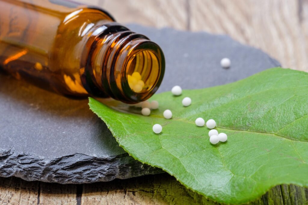
Homeopathy – what is it? Does it really work? This is a hugely debated question within the pet industry as well as the human medical space. With little scientific evidence in the past, surprisingly, more and more evidence is being released in more recent years to suggest that homeopathy is in fact beneficial! Here at MPN, we aim for a holistic approach throughout our consultations; and as part of that, we may suggest speaking to a qualified homeopath alongside our help for your pet! Read on to learn more about homeopathy! How Are Homeopathic Remedies Made? Homeopathic remedies are totally natural, and are based on extraordinarily diluted extracts from plant, mineral and animal with medicinal properties when used in such weak dilutions. Due to the nature of some of the plants from which these remedies are based on being toxins as a whole plant, it is imperative that you purchase your homeopathic remedies from a licensed homeopathic pharmacy. Our favourites are Helios Homeopathy, and Ainsworths Homeopathic Pharmacy. Remedies used in pets, typically come in the form of pillules – these are small balls made of sugar, coated in the remedy. Other forms remedies are available in include tinctures (also popular for pets, especially those from the brands ‘PhytoPet’, and ‘Dorwest Herbs’), granules, powders, larger pills, and creams/ointments. The vast majority of remedies are made through repeated levels of dilution. Whichever plant or animal the remedy is based on, will be cut down to extract only the relevant part of the plant (for example, the remedy Fragaria, often used for pets to aid dental health, is based on the ripe fruit of the wild strawberry, so the leaves will be removed from the plant to begin with). This raw material will then be crushed and dissolved in either water or alcohol, depending on the nature of the raw material. A process called ‘succession’ is then used to dilute the dissolved material – during this process, it is shaken vigorously with water or alcohol. This process is repeated a large number of times until the intended level of dilution is reached. Once the desired dilution is reached, the product left is the active ingredient in the homeopathic remedy, and is used to further create the remedy sold to the consumer, in the desired presentation. The remedies each come in a variety of strengths. The strength required for an individual depends on the symptom it is aiming to support. Typically, in the UK, you will notice remedies using the centesimal scale in terms of strength; so your remedies will usually be ‘6C’, ‘30C’ or ‘200C’, though you may also see the decimal scale used which would be displayed as ‘6X’ or ‘30X’, as examples. Those using the centesimal scale are more potent than those using the decimal scale. As an example, let’s look at the difference between a remedy that is 30C, and one that is 30X. The 30C remedy would be more dilute than the 30X remedy because the 30C remedy has been diluted in a ratio of 1:100 (1 part substance containing the raw material, 100 parts water/alcohol) 30 times, whereas the 30X remedy would have been diluted in a ratio of 1:10 (1 part substance containing the raw material, 10 parts water/alcohol) 30 times, meaning the 30C remedy is much more dilute than the 30X remedy. The usual ‘go to’ for pets is a 30C potency. 3C or much higher potencies may be advised by a homeopath in certain circumstances. How Does Homeopathy Work? When we look at the mechanism of how homeopathy works, we can’t just say ‘your dog has this problem, this is the remedy for that problem’ like your vet may with pharmaceuticals, we have to look more into the pet as a whole; their symptoms, diagnosis, and emotions all partly determine which remedy you may need to consider for your pet. Speaking with a homeopath is incredibly important as getting the correct remedy is important, especially when there are two or more which may fit your dog’s symptoms! In homeopathy, the remedy advised by a homeopath would depend on the ‘Law of Similars’; whereby ‘like cures like’. The pattern of symptoms caused by the original source a homeopathic remedy is diluted from, makes it the best remedy to treat the symptoms your pet is displaying. Findings Here Findings Here Findings Here What Conditions Can Be Supported With Homeopathy? There’s definitely no specific number we can answer this question with! The list of symptoms you can support with homeopathy is endless; we’d be here all year if we were to list them all! Almost every aspect of emotional and physical wellbeing can be supported through the use of homeopathy. Using the Homeopathic Materia Medica, you will be able to match symptoms to a remedy. The Materia Medica. The Materia Medica is a book describing the history of homeopathic remedies, and gives a detailed list of remedies, and which remedies match with which symptoms. The Helios Homeopathy Pet Kit includes it’s own Materia Medica which explains how to use the remedies, and lists possible uses for each remedy that you may encounter a need to use. An online complete Media Medica, written by homeopathic physician William Boericke in 1901 is available here. Popular Remedies in Pet Healthcare There are thousands of homeopathic remedies available, but there are some more commonly used in pet care. Here are six of the most popular remedies used in pets: Arnica: this is a very popular, and useful remedy, often given to those following an injury or operation, as it is often prescribed for bruising, sprains and strains, and arthritis. Studies suggest effects of Arnica can be comparable to those of anti-inflammatories. Findings Here Findings Here Pulsatilla: this remedy is typically used to reduce the symptoms associated with phantom pregnancy in pets. It can also be used to aid tender gums, throat and mouth. One study shows the use or pulsatilla in 4 dogs with eye issues and separation anxiety was paramount
Frequent readers of our blogs will know we aid pet owners with the vast majority of health complaints in their beloved furry family members. One of the health complaints we see in puppies is ‘Puppy Strangles’. Being a life threatening disease, owners of puppies suffering with this are right to be concerned! Read on to find out more about Puppy Strangles, what it is, how it is caused, diagnosed and treated, and how we can support healing naturally. Puppy Strangles is a rare skin disease affecting puppies, often referred to as Juvenile Cellulitis, lymphadenitis, or sterile granulomatous dermatitis. The disease can be life-threatening if left untreated, so acting fast and seeking veterinary care is essential. While Puppy Strangles has been seen in dogs of up to 4 years of age, it is most commonly seen in puppies of 6 months and under. Symptoms and Causes There are a variety of symptoms associated with Puppy Strangles. Some symptoms you may see in a puppy with this condition are: Sudden facial swelling: the swelling you may expect to see will show rapidly, and can usually be seen first around the muzzle and eyelids. Eventually, the lip area, and the underside of the chin may be affected in a similar way. Lesions on the skin: in some cases, lesions may also form on the skin, primarily around the face, but also seen in some cases around the paws, anus and vulva. Swollen lymph nodes: the lymph nodes located under the chin and in the neck will swell, looking like small, soft lumps. This is one of the biggest signs of Puppy Strangles. Reduced appetite: as your puppy will be significantly unwell, their appetite may reduce. Lethargy and fatigue: your puppy’s immune system will be heavily impacted, and will be taking a huge amount of energy, resulting in lethargy and fatigue. Lameness: due to the inflammation in the body, and lack of available energy, your puppy may show signs of lameness. Fever: the body temperature will rise due to inflammation. Behavioural changes: many puppies display low mood behaviours when severely unwell. When looking at causes of Puppy Strangles, there are a few avenues under exploration, but the overall cause is largely unknown at present, and it is deemed ‘idiopathic’, meaning the cause is unknown, and it happens spontaneously. There are some suggestions that this condition has aspects of an autoimmune disease, whereby the dog’s immune system malfunctions and causes the body to attack it’s own skin. There is also a chance that the disease has hereditary predispositions, meaning the risk is passed from generation to generation. There may also be breed predispositions for puppy strangles, as the breeds most commonly affected appear to be dachshunds (all sizes and coat types), Gordon Setters, and Golden Retrievers. Findings Here Findings Here Findings Here Diagnosis Diagnosing puppy strangles can be a complex task. There are various methods used, some vets will use multiple methods, some will use one or two. The diagnosis methods include: Physical examination: as the visible symptoms for Puppy Strangles are so noticeable, the first part of diagnosis is a physical examination of the skin. the vet will look at the puppy’s overall condition and look for lesions etc. Checking the puppy’s medical history: the age, breed, and family history of the puppy will be analysed. Skin biopsies: samples of skin from affected areas may be taken. A full laboratory report will be written regarding the biopsies after analyses under a microscope. This is the most conclusive form of diagnostics for Puppy Strangles. Skin lesion cytology: in some cases a small sample of tissues from a skin lesion will be taken and analysed under a microscope to rule out infection in these areas. Skin scrapes: the veterinarian may take scrapings of the top layer of skin, and take a look at these under a microscope. This will enable them to rule out possible mites or fungal infections which can present similarly to Puppy Strangles. Hair plucking analysis: much like skin scrapes, examining plucked hairs under a microscope will rule out parasitic or fungal burdens. Blood panels: a full blood panel will often be run, which will show any potential discrepancies which could form a different diagnosis, and to check the white blood cell count of the individual. A classic symptom of Puppy Strangles is an elevated white blood cell count. In cases where a veterinarian suspects a secondary infection in an individual, they may also perform fungal or bacterial cultures to rule out further infection. Findings Here Findings Here Findings Here Conventional Treatment As Puppy Strangles is immune mediated, the immune system needs to be suppressed to treat the disease. If the immune system is not suppressed, the body will continue to attack itself, causing the condition to worsen, and the puppy’s life to be at risk. An immunosuppressive drug (usually a corticosteroid such as prednisone) will be administered to your puppy, daily over the course of a few weeks in order to combat the disease. After the initial few weeks, the dose will be gradually tapered as your dog will need to gently wean off it. Due to the risk of infection in skin lesion sites I those with Puppy Strangles, it is common for antibiotics to also be prescribed by your veterinarian as a precaution. Anti-inflammatory medication may also be prescribed to reduce inflammation in the body, caused by the disease. It is imperative that you follow your veterinarian’s advice, but you may wish to use complementary support alongside treatment. Findings Here Findings Here Findings Here A Natural Approach to Puppy Strangles As always, our approach is often supported by veterinary care, so we tend to look at this as a complementary approach as opposed to alternative. Side effects of short-term use of the aforementioned drug protocol can include increased appetite, thirst and urination, as well as reduced gut microbiota diversity and microbiome health, and increased lethargy. Complementary support can go some way to reducing these side effects. Here are some
Pet dental health is so important for all! At My Pet Nutritionist, we love to educate owners on all aspects of pet health and husbandry. Many pet owners come to us when their pet’s oral health needs improvement; dental surgery is often required, but what do you do if your pet needs dental surgery? This blog post aims to help you understand why a pet may need dental surgery, what to do before surgery, after surgery, and ongoing to reduce the risk of needing further dental surgery. Why Might Your Pet Need Dental Surgery? There are many reasons a dog or cat may need dental surgery. Some mild conditions or cases of dirty teeth may be resolved at home, but many pets require professional help to avoid severe health complications caused by very poor oral health. Here’s some of the most common reasons a pet may need dental surgery: Periodontal Disease: pets with periodontal disease often end up going undiagnosed for a long time, causing the disease to develop. There are various symptoms of periodontal disease including bleeding gums, bad breath, receding gums, tooth loss, pus around the teeth, tooth discolouration and formation of epulis. If left untreated, periodontal disease can be life threatening, as bacteria emanating from the mouth enters the gut via the bloodstream. Organs commonly affected by periodontal disease are the kidneys, liver and heart. If your pet has diabetes, you may also find blood sugar regulation more difficult. Fractured Teeth/Jaw: if your pet manages to fracture their jaw or teeth, this will require dental surgery. Generally speaking, fractured teeth will be removed; some cases may be offered root canal surgery, but it is usually easier to have the tooth removed. Fractured jaws can require more extensive surgery, and come with a longer recovery process. Retained Juvenile Teeth: some young dogs require tooth removal surgery due to retained juvenile teeth. This is when the adult teeth have grown in front of, behind, or next to the puppy teeth, but the puppy teeth have not detached from the gumline, leaving two sets of teeth. Most puppies won’t require this intervention, but some may. Oral Tumours: while rare, oral tumours can happen in dogs at any age, but particularly in older dogs, or dogs with poor dental hygiene in general. Dental surgery is usually recommended to remove the tumour, and surrounding tissues if the margin is available. Some oral tumours will require removal of parts of the jaw. Cleft Palate: in rare cases, a pet may suffer with a cleft palate, which is a congenital defect whereby the roof of the mouth has failed to form properly. This lack of closure results in an opening being left between the mouth and the nasal cavity. Dog breeds most susceptible to this are the brachycephalic breeds (pugs, French bulldogs, English bulldogs, Boston terriers), and cat breeds most prone to this condition are the Siamese, Persian and Savannah cats. Findings Here Findings Here Should I See A Specialist? We recommend seeking help from a veterinary dental specialist! While traditional vets are amazing people, worth their weight in gold, when your dog is undergoing dental surgery we recommend seeking help from a veterinary dental surgeon. The tissues in the mouth are very delicate, and while traditional veterinarians learn about oral health, veterinary dental surgeons are highly skilled in their specialist area, with far more in-depth knowledge when it comes to oral health and disease. Post Diagnosis Preparation Once you have received a diagnosis for your pet’s dental issue, and are awaiting a date for their dental surgery, your veterinarian will most likely give you a short list of things to do between diagnosis and surgery. This list may include some of the following: Feed a softer food: while waiting for surgery, your pet is likely to require a soft food. Softer foods will put less strain on the gums and teeth themselves, which will therefore decrease the risk of pain in these diseased or damaged areas. Many pets lose their appetite when their mouth is painful, which may also create a negative association with food. Some pet owners choose to feed a soft, high quality, high meat content wet food, others may choose to feed a cooked food with added bone broth to soften it. Avoid chews: as chews are generally hard or chewy in texture, allowing your pet access to these may cause pain or further aggravate the affected area. Remove unsuitable toys: some of the toys your vet may recommend removing from your pet’s reach include knotted toys, tug toys, solid toys and balls. Avoiding use of these toys will help reduce discomfort for your pet, and reduce the risk of further damage prior to surgery. Keep these toys out of reach until oral wounds are healed. Antibiotics: depending on your pet’s diagnosis, your vet may recommend a course of oral antibiotics to reduce risk of infection in the affected area, or to treat infection that may already be present. As with any course of antibiotics, it is important to finish the whole course prescribed, unless advised otherwise by your veterinarian. Painkillers: as dental health problems usually cause discomfort or pain, your vet is likely to prescribe painkillers in the time between diagnosis and surgery. Ensure your pet is given these, and look out for any changes in behaviour which may suggest worsening of pain. Practice crate training/calmness: some pets my require a complete rest period after surgery, depending on the procedure required. As activity increases blood flow to the gums, you will most likely need to keep your pet calm following surgery to allow the surgical site to heal. Ensuring your pet is both comfortable in a crate, and able to relax without being overstimulated by normal household life is incredibly beneficial to their post operative period. The more comfortable they are in these scenarios before their surgery, the more relaxed they will be during recovery, which aids the speed of recovery. Findings Here Preparation for the Night Before/Morning
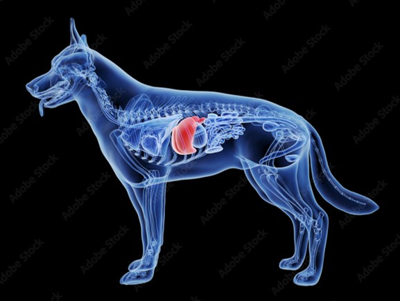
Here at My Pet Nutritionist, we love to take an in-depth look at all organs and systems within the body in an approach to overall health and well-being in pets. As part of our mission to help pet owners keep their pets in the best health possible, some of our blog posts are designed to target one specific part of the body – this blog post take a deep dive into everything liver! Learn what the liver is, what it does, common complaints we see within our customer base, and how to support the liver through diet and lifestyle changes! Liver Structure The liver is the largest internal organ in the body. It is found inside the ribcage of dogs and cats, just below the diaphragm, and while it is a rough triangle-type shape, it is soft, and takes the impression of the surrounding organs including the gallbladder, stomach, pancreas duodenum and the right kidney. It is a large organ, and consists of 6 lobes. Each lobe is made up of many ‘hepatic lobules’, and each lobule holds a large number of blood filled, sac-like cavities called sinusoids. Due to this structure, the liver is a spongey texture, and is able to hold large amounts of blood, which gives the organ a deep red colour. There are two ways fluids travel into the liver; both major blood vessels! The first, known as the Portal Vein takes nutrient rich blood from the gastrointestinal tract into the liver, however this vessel also brings the not-so-beneficial products of chemicals and drugs into the liver too! The other major vessel is called the Hepatic Artery, which delivers oxygen rich blood from the heart and lungs, into the liver. Just like routes into the liver, there are two ‘exits’ too! Hepatic Veins carry blood out of the liver, whereas the Bile Ducts take bile (a yellow coloured digestive substance made in the liver, and stored in the gallbladder) out of the liver, and into the gallbladder. The liver can also self-regenerate if it is compromised through trauma!’ Findings Here Findings Here Findings Here What Does the Liver Do? The liver has many major functions within the body, affecting various aspects of health. Let’s take a look at these: Bile Production: the liver plays an enormous role in digestion. It produces bile, a yellow coloured digestive juice, which is absolutely essential for the digestion of fats in the small intestine and nutrient absorption. Metabolism: the liver’s role in metabolism includes breaking down protein, fat and carbohydrates in order for the body to be able to use them as energy. The energy gained through this metabolic process is stored in the liver as glycogen which is released when required. Vitamins and minerals are also stored by the liver, and inactive forms of these are made active. Blood Detoxification: the liver is the body’s very own toxin removal system! When there are unwanted substances in the blood, the liver removes them from the body. As part of this function, the liver also breaks down any medications in order to eliminate the toxins. Filtration from digestive tract: not only does the liver filter the blood for toxins, but it also filters blood from the digestive tract. Blood Clotting: the liver is home to a variety of proteins which clot blood. These proteins include various coagulation factors, fibrinogen, and prothrombin. Immune aid: the liver plays a role in the immune system too! Some essential proteins and enzymes used in the immune system are produced in the liver. Blood storage: finally, due to the liver’s texture and storage capacity in the sinusoids, it is excellent for storage of blood which it releases if required. Findings Here Findings Here Findings Here Common Liver Complaints Many of our customers come to us for help with their dog’s liver problems. We have helped to support those with a wide range of liver complaints over the years. Some of the most common complaints we see are: Liver Disease: liver disease has many possible underlying causes including genetics, hepatitis, liver tumours, infections or toxin exposure, including from medications and some plants. Liver disease can have a lot of knock on effects on the body including disturbances in filtering toxins, difficulties storing nutrients, and reduced digestive capabilities. Symptoms include increased thirst (polydipsia) and urination (polyuria), vomiting, reduced appetite, rapid weight loss, and jaundice (yellowing of the skin). Diagnosis is sought via a combination of blood test, imaging, and sometimes biopsy of the liver to discover the underlying cause. Liver Shunts: clinically, these are referred to as portosystemic shunts. Dogs with liver shunts have abnormal blood vessels which bypass the liver, meaning the liver cannot perform it’s job of filtration of, or nutrient metabolism from the blood efficiently. Liver shunts can be congenital defects (present from birth), or caused by some types of liver disease. Symptoms of liver shunts include vomiting, diarrhoea, hypersalivation, stunted growth, reduced coordination, seizures, and behavioural changes. Blood tests and imaging (including ultrasound and CT scans) are used to diagnose a liver shunt. Hepatitis: this condition is a chronic inflammatory condition in the liver, often caused by infection, toxin exposure, or immune dysfunction. There are various types of hepatitis, the most infectious one being Adenovirus Type 1 which is potentially life-threatening! Fever, lethargy, vomiting, jaundice, and enlarged liver are all common symptoms of hepatitis. Lifelong liver damage can occur depending on the severity of infection. To diagnose this condition, imaging, blood tests and often liver biopsies are performed. Liver Cancer: liver cancer can be classed as ‘primary’ in which the abnormal cells originate inside the liver itself, or ‘metastatic’ where the abnormal cells spread from other parts of the body. Reduced appetite, weight loss bloating and lethargy are the initial symptoms one can expect, and these may not be clearly linked to liver cancer. More advanced symptoms of liver cancer are those of liver failure. Biopsies, imaging and blood tests are used to diagnose the condition. Acute Liver Failure: the term ‘acute’ refers
Here at My Pet Nutritionist, we know that a healthy gut microbiome is the most important aspect to keeping your dog healthy all round! There is so much to learn about the gut microbiome – what it really is, what it consists of, what other parts of the body it interacts with, and what happens when it’s not in tip top condition. Many of our readers will notice that most of our blogs mention the microbiome, so this blog post is a full round up of everything gut microbiome related, in one easy read! What is the Gut Microbiome? Every dog has a gut microbiome. It’s one of the most important parts of your dog in terms of digestion and overall health, from joints to skin, to the brain and immunity. The microbiome is a community of microbes, including good and bad bacteria, viruses, and fungi. The gut microbiome is very carefully balanced. Some of the microbes found in a healthy gut microbiome are ones we would typically class as ‘bad’ which have health conditions associated with them, including bad bacteria such as Salmonella, and Clostridia. Viruses and Fungi are typically classed as ‘bad’ microbes too, but are still part of a healthy microbiome. Good bacteria such as Lactobacillus, Bifidobacterium and Enterococcus are all found in abundance in the healthy canine gut microbiome – these beneficial bacteria are found in high enough numbers to outweigh the negative impact of the bad microbes found in the gut. A healthy gut microbiome means the ‘good guys’ outweigh the ‘bad guys’. Health problems begin to emerge when the gut microbiome is knocked out of balance, allowing for gut dysbiosis to occur. Gut dysbiosis can lead to a host of health problems including reduced skin and joint health, increased digestive issues, and prominent or mild negative behavioural changes. Findings Here Findings Here Interactions Between the Gut Microbiome and the Rest of the Body The health of the gut microbiome is a huge part of overall health – but why is this? You may be wondering how gut health could impact so many other parts of the body, so let’s take a closer look at these! We can look at the gut as a ‘roundabout’. Roundabouts have multiple exits – these ‘exits’ stemming from the gut are collectively called Axes; and these link to other parts of the body to form a two-way pathway/link from the gut. The main axes are: The Gut-Skin Axis: this pathway connects the gut to the skin, and the skin to the gut. If we start at the gut and work our way to the skin, we can see that poor gut health can cause or exacerbate skin conditions. Going to other way; from skin to gut, we unfortunately have the risk of gut microbiome damage caused by external exposure to toxins, whether they’re from laundry detergents, household cleaning products, external flea and tick medications, or other environmental toxins. The Gut-Joint Axis: this pathway connects the gut to the musculoskeletal system, and the musculoskeletal system to the gut. An imbalance in the gut microbiome can be connected to poor joint health, especially in cases of osteoarthritis due to the increase in inflammation around the joints. In a poorly balanced gut, there is often an increased risk of pro-inflammatory cytokines and inflammatory metabolites being produced, which are the main cause of poor joint health in dogs. The Gut-Brain Axis: this is the bidirectional pathway between the brain and the gut. This pathway has connections both physically, and chemically. In terms of physical connections, the Vagus Nerve is the main avenue for carrying signals between the two locations. Chemically, the healthy gut produces neurotransmitters and hormones, which move between the brain and gut. Mood, response to stress, and digestion are three of the main roles of the gut-brain axis. It is also important to note that around 70-80% of the immune system is found in the gut! Keeping the gut microbiome healthy, and perfectly balanced means the immune system, and therefore overall health of an individual remains stable. Findings Here Findings Here Findings Here Findings Here What Happens When the Gut Microbiome is Unhealthy? When the Gut Microbiome is unhealthy, a host of health conditions can occur. As previously mentioned, an unhealthy gut microbiome is unbalanced; and this condition is called Gut Dysbiosis. Gut Dysbiosis occurs when the ‘bad microbes’ outweigh the ‘good microbes’. Here’s some health concerns that we see a lot in those with an unhealthy gut microbiome: Food sensitivities: when the gut microbiome is unbalanced, food sensitivities are common due to increased gut permeability; known as Leaky Gut. When the gut integrity is poor, food particles leak into the bloodstream which encourages the body to create an immune response, presenting as food sensitivities. Yeast: yeast cells are naturally present in the gut; they’re called Candida. When Candida is present, and there’s not enough of the good microbes to keep these in check, a yeast infection will often occur. Symptoms include rust coloured paws, ears and groin, cheesy smelling feet, and itching among others. Digestive problems: digestive upset including diarrhoea and vomiting is common in those with an unhealthy gut microbiome, due to the abundance of ‘bad’ microbes and a lack of ‘good’ ones to keep the effects of the bad ones at bay. Many dogs also suffer with Small Intestine Bacterial Overgrowth (SIBO) which contributes to these symptoms. Bad breath: a lot of owners assume their dog’s bad breath is caused by poor dental hygiene, however this is often not the case. Bad breath can emanate from the gut, in those with poor gut health. When the gut microbiome is unbalanced, the overgrowth of harmful bacteria in the gut results in the production of VSCs; Volatile Sulphur Compounds, which cause bad breath. Inflammatory Bowel Disease (IBD): when the gut microbiome is unbalanced, inflammation occurs as a result. This inflammation often leads to chronic inflammatory conditions such as IBD. Other gastrointestinal conditions occur due to this too, however IBD
Here at My Pet Nutritionist, we use a combination of healthy diets tailored to your pet’s needs, alongside beneficial gut-friendly herbs, and nutraceuticals. These supplementary additions often come with hosts of benefits, when used in the correct way, for a specific purpose. We often get asked about the various herbs and nutraceuticals we recommend in various blog posts and in our recipes and supplements – you can learn more about some of these here! What Are Gut Healing Herbs? Herbs high in mucilage are often known as the ‘gut healing herbs’. These herbs are usually considered to be a source of prebiotics – food for probiotics; however these herbs have another very important function when it comes to gut health! Mucilage is a soluble dietary fibre, which produces a mucus-like substance which lines the gut wall. When the gut wall is lined with this slimy textured substance, it forms a protective barrier to reduce the risk of further gut damage, and allow the gut to heal. Inflammation is then reduced due to lack of irritation. Mucilage has a fantastic soothing effect on the digestive tract too – those with acid reflux, or having recovered from illnesses like kennel cough which may cause throat irritation may find a powdered form best, due to the soothing effect of the mucilage in these herbs. As an additional benefit of these popular gut healing herbs, the mucilage can increase digestive performance resulting in better stools; this is due to the mucilage adding bulk to the stool (reducing constipation), aiding water absorption, and increasing gut motility. Some studies also suggest that mucilage rich herbs may reduce the build up of cholesterol, and aid the regulation of blood sugar levels. Frequently used mucilage herbs include slippery elm, marshmallow root, and deglycyrrhizinated liquorice root – we will look closer at these later. Findings Here Findings Here What Are Nutraceuticals? Nutraceuticals are components of foods that bring benefit to health. You may often come across plant based supplements for pets; these are packed full of nutraceuticals! Functional foods are also classed as nutraceuticals. Many nutraceuticals enhance basic nutrition, whereas some provide medicinal properties. Some benefits of using carefully selected nutraceuticals include improved overall health, prevention of chronic disease, increased longevity and all round structural support for the body. The term ‘nutraceutical’ is a combination of the words ‘nutrition’ and ‘pharmaceutical’ – meaning they share the same properties as pharmaceuticals, but are acquired through nutrition Functional herbs, antioxidants, probiotics, vitamins and minerals are all examples of the categories most commonly encountered in the world of pet nutrition. We will delve into these categories in terms of nutraceuticals later. Findings Here Findings Here When Should We Use Gut Healing Herbs and Nutraceuticals? A question we often hear at My Pet Nutritionist, is “should we be giving any supplements?” – and our answer is always “if your pet could benefit from a carefully selected supplement, then absolutely!”. It is important not to needlessly add lots of different nutraceuticals and gut healing herbs to your dog’s diet, unless they require/could benefit from it. When it comes to giving mucilage herbs, these can be incredibly beneficial in situations where gut damage is apparent. Situations a mucilage herb may be beneficial include: Allergies: due to an immune malfunction Intolerances: due to gut damage or leaky gut syndrome Following vaccines, worm or flesa treatment: these products are very damaging on the gut, so gut healing may be necessary. Acid reflux: soothes the digestive tract and reduces the risk of acid reflux Upset stomach or constipation: these herbs help to rectify loose or solid stools when needed. Great to have in the first aid cupboard! When looking at nutraceuticals, we need to look at the symptoms your dog or cat is displaying, alongside gaining a diagnosis from your veterinarian. Some types of illnesses you may use nutraceuticals for include: Joint issues Skin issues Digestive issues Cognitive health issues (anxiety etc) Vision issues As brilliant as nutraceuticals can be as part of your pet’s health regime, many do come with some contraindications with pharmaceuticals your pet may need. Contraindications are negative interactions with drugs – these interactions can often cause downregulation of the drugs themselves, leaving your pet vulnerable to disease progression. An example of this is curcumin – the active compound in turmeric. It is fantastic for joint support/arthritis, cardiovascular function, reducing inflammation, eradicating free radicals, immune support, cognitive ability and cancer prevention. The reduction in pain in those who take curcumin is vast; however some individuals may require pain pharmaceutical pain relief for their condition. Curcumin is known to downregulate the effects of some pharmaceutical analgesics, as well as various other drugs. It is always best to check with your veterinarian before starting a new nutraceutical, if your pet is on pharmaceutical drugs. Findings Here Findings Here Popular Gut Healing Herbs The three most popular gut healing herbs can all be found inside our Gut Guardian supplement, along with probiotics and chamomile. Lets take a closer look at these mucilage herbs! Marshmallow Root: this is a sweet smelling and tasting herb derived from the Althaea officinalis plant. When combined with water, it immediately forms a thick, slimy substance due to it’s high mucilage content. Traditionally, marshmallow root is used in cases of digestive or respiratory issues, and also topically to soothe skin issues. As well as being high in mucilage, it also contains high amounts of antioxidants, anti-inflammatory properties, and antibacterial properties. slippery elm: like marshmallow root, slippery elm produces a slimy substance when combined with water. It originates from the inner bark of the slippery elm tree (Ulmus rubra), and is often used to treat digestive issues, and sore throats by lining the oesophagus. DGL (deglycyrrhizinated liquorice): this herb increases mucous production in the body, which helps line the stomach, reducing the risk of harm caused by acid. DGL is thought to have properties to treat ulcers too! Findings Here Findings Here Findings Here Popular Nutraceuticals There are many nutraceuticals used in the
In part two of our focus on the diet of a senior dog, we will look further into some specific nutritional categories bringing fantastic health benefits to your senior dog. We will also go through our recommendations on what to feed your senior pet, including specific nutrients and supplements you may wish to consider! Read Part 1 here! Specific Beneficial Nutrients There are a number of macro- and micronutrient categories senior dogs would benefit greatly from to slow the ageing process down, and reduce age-related inflammation. The two main categories to analyse here are Antioxidants, and Omega Fatty Acids. Antioxidants: these are one of the most important categories of nutrients to feed your senior dog (and are highly beneficial at any age!) due to their action against oxidative stress. As dogs age, oxidative stress becomes more proliferative; it’s a natural part of ageing! Oxidative stress is caused by ‘free radicals’ – unstable oxygen molecules missing an electron. Free radicals damage all parts of the body from organs to joints, causing joint degeneration, cancer, altering DNA, neurodegenerative diseases, adverse alteration of fats and proteins, and other diseases like diabetes, so it is incredibly important to combat these cells as best you can. Antioxidants tackle and remove free radicals from the body, leading to less oxidative stress, and therefore slower ageing. Some excellent sources to think about including in your senior dog’s diet include blueberries, strawberries, artichoke, goji berries, red cabbage, kale, ginger, spinach, parsley, pecans (in small amounts due to fat content), fresh garlic (avoid in Japanese breeds) and rosemary (avoid in epileptic dogs). Omegas Fatty Acids: this group of fats are another incredibly important category of nutrients to include in your senior dog’s diet, specifically Omega 3. The two ‘main’ omega fatty acids are Omega 3 and 6 – both are essential. Omega 9 is present in the diet too, but doesn’t add as many benefits to the dog’s health as Omegas 3 and 6. Omega 6 fatty acids called Linolenic Acid (LA) and Arachidonic Acid (ARA) are inherently inflammatory. The only non-inflammatory Omega 6 is Gamma-linolenic Acid (GLA). Omega 6 brings many benefits to the health of the dog despite it’s inflammatory properties, including control of hormones, however as it is naturally in abundance in our carnivorous pets’ meat based diet, we need to balance the inflammation out! This is where Omega 3 comes in! Omega 3 includes Eicosapentaenoic Acid (EPA), Alpha-linolenic Acid (ALA) and Docosahexaenoic Acid (DHA), and is inherently anti-inflammatory. The anti-inflammatory properties of Omega 3 Fatty Acids balance out the inflammation caused by Omega 6 Fatty Acids. EPA is the main inflammation reducer, while ALA aids immune and heart health, and DHA aids cognitive health and development. Some excellent sources of Omega 3 to include in your dog’s diet include fatty fish and fish oils, algal oil, flax and chia seed, oysters and eggs. You can read more about choosing an omega supplement for your pet here! Findings Here Findings Here Findings Here What Type of Food Should I Feed? The dog food market really is a minefield! There are many options of different food types, each claiming to be the best! Our ethos here at My Pet Nutritionist is to feed as fresh food as possible! Minimal processing is a major factor in healthy ageing and longevity. In this article, we are going to take a closer look at dry food, fresh cooked food, and raw food. Dry food is not a diet we would generally recommend, especially not for a senior dog! But why? High carb: carbohydrates can have an inflammatory effect on our pets. Dry foods are typically between 30 and 60% carbohydrate – sometimes even higher! Manufacturing process: these foods undergo a huge amount of processing. During the cycle of production from raw ingredient to bag, these foods are subject to up to 4 separate high-heat processes, each time reducing nutritional quality. Synthetic vitamins and minerals: our senior dogs may not be able to utilise the synthetic vitamins and minerals often sprayed onto the dry food at the end of the manufacturing process, deeming them useless. Glyphosate: due to the extreme manufacturing process, glyphosate production occurs. This is the main ingredient effective against killing weeds, but it is linked with cancer and damage of the gut microbiome. Mycotoxins and Aflatoxins: mycotoxins are chemical compounds caused by mould and can lead to vomiting and seizures. Aflatoxins are also caused by mould in food and can lead to cancer and liver damage. Storage mites: before bagging, dry foods are often stored in large hoppers – and even after bagging, the shelf life is very long, encouraging storage mites which can trigger allergies. Advanced Glycation End Products: shortened to AGEs, these harmful chemical compounds are produced during manufacturing, and a linked to cancers, inflammation, oxidative stress and premature ageing. Why do we advocate for a fresh cooked, or raw diet? Which is best for senior dogs? Fresh food is minimally processed, meaning the harmful compounds listed above do not form, reducing the risk of inflammation, oxidative stress and other disease in the body. As the ingredients are still in their whole, natural form they are nutrient dense, and provide the body with excellent nutrition! Another major benefit to fresh food, is that it is high in moisture – moisture in the diet is essential for kidney and gut health, both of which are known to worsen during the ageing process! As the gut motility of senior dogs can reduce, and other changes in the gut naturally occur, many seniors cope best with fresh cooked food as opposed to raw food – though some do still tolerate raw food! We have an extensive array of well formulated and balanced meal recipes for dogs on our website! Our Recommendations There are various aspects of diet we actively encourage owners of senior dogs to include in their dog’s feeding regime. Some may benefit from a more tailored 1-2-1 approach with one of our consultants, however our general
Here at My Pet Nutritionist, we help pet owners help their beloved dogs and cats with all aspects of health and nutrition. Those we consult for include all ages, from puppyhood right through to old age. Our golden oldies deserve just as much love and care as their younger counterparts! As a dog ages, their dietary needs may change – but how? In this bumper 2-part blog, we take a look at the dietary changes your senior dog may need to stay happy, healthy and pain free! Is My Dog a Senior? This is a question we here ever so frequently! Is your dog a senior? Is there a specific age your dog becomes a ‘senior’? what are the signs your dog is becoming a senior? Many processed dog food manufacturers will class a senior dog as ‘7 years plus’ – however we know there are so many contributing factors to a dog becoming a ‘senior’, and this isn’t always at 7 years of age! Factors affecting a dog’s ageing include: Breed: generally speaking, larger breeds will hit their senior years far earlier than smaller breeds. Breeds with predispositions to health conditions may also reach senior years earlier than those without. Genetics: a dog’s family genetics/history can affect the age they reach senior years. Medical history: if your pet has had any health complications growing up, any medical reactions, infections etc, these may cause senior years to come sooner. A dog’s neutering status, or the time they were neutered may also contribute to ageing. Dietary History: a dog fed a gut-friendly diet, full of natural nutrients, with minimal processing for life is more likely to reach senior years later than those fed an ultra-processed diet full of synthetic micronutrients. Ultra-processed dry foods also tend to have a high content of pro-ageing substances called Advanced Glycation End-Products (AGEs) which cause faster ageing. Exposure to toxins: toxin exposure can cause disruption in the endocrine (hormone) system, and cause gut stress which usually comes hand in hand with life shortening conditions, causing senior years to approach quickly. These toxins can be anything from flea and worm medications to environmental toxins both in the home and out and about. Behavioural History: even your dog’s behaviour can have an effect on longevity! This may seem like an odd link, but behavioural stress can have an effect on physical stress, and vice versa. The pathway between the brain and gut, known as the gut-brain axis is at play here! Physical stress on the body, caused by behavioural stress, will speed up the process of ageing. Signs that your dog is entering their senior years include: Weight loss Reduced hearing Reduce eyesight Increased fatigue Less willingness to exercise Reduced cognitive ability Stiffness Reduced appetite Increased water consumption Development of lumps and bumps Lack of balance and stability. Of course, as your dog enters the early staged of senior-hood, these symptoms may be mild, and your dog may only display a few. As your dog progresses through their senior years, more symptoms may appear, or existing symptoms may worsen. Nutritional Needs of a Senior Dog As your dog ages, various changes occur throughout their body which require some nutritional tweaks to ensure optimum health continues. These changes include various internal systems in the body such as: Gut health: the gut of an ageing dog can be subject to various changes affecting gut motility, nutrient absorption and processing. Digestive capabilities often occur due to gut degeneration. Brain health: ageing canines are often subject to cognitive decline. Joint health: one of the major pathways from the gut is the gut-joint axis. When the senior dog’s gut is compromised, joint conditions can become more prominent. Senior dogs generally require more joint support than younger dogs. Dental Health: dental conditions like gingivitis, tooth decay, periodontal disease, tooth fractures and tooth resorption are often seen in older dogs. Kidney Health: kidney disease is one of the more common age-related diseases we often hear about here at My Pet Nutritionist, as older dogs are more susceptible to Chronic Kidney Disease; a progressive disease. Heart Health: as the heart is a muscle, over time it can begin to fatigue. Statistically, around 75% of senior dogs suffer some sort of heart disease! Findings Here Findings Here Findings Here Findings Here When it comes to nutritional composition, we need to look closely at the macronutrients and micronutrients in your dog’s diet; both quantities of, and types/sources of. Macronutrients are the main nutrients our bodies require in larger amounts; let’s take a look these requirements for a senior dog: Protein: The building blocks of protein are called Amino Acids. Amino Acids are essential for pretty much everything in the body to form and function normally, including muscles, tendons, ligaments, cartilage, hair, nails and skin. Protein is also used in the endocrine system, to enable the healthy production of hormones throughout the body. As the dog ages, protein deficiencies become more common, which can lead to absorption issues and muscle degradation which ultimately affects mobility. A meat based, moderate-high protein diet is advisable for senior dogs. Fat: as dogs become less active due to the natural ageing process, a low fat diet may be advisable. Feeding a high fat diet when activity levels are waning may cause weight gain which puts pressure on joints and other parts of the body. The type of fat included in the diet also makes a difference! Saturated fats are the type we ideally do not want much of, however Omega 3 Fatty Acids are known for their anti-inflammatory properties, and are an important part of s senior dog’s diet! More on this later. Carbohydrate: ‘complex’ carbs which are high in fibre may be a great addition to your pet’s diet, to improve digestion, regulate metabolism and help maintain the immune and nervous systems. Higher fibre carbohydrate options include kale, broccoli and leafy green vegetables. Other ‘complex’ carbs that are ok to be fed in small amounts include sweet potato, pumpkin, banana, berries
Here at My Pet Nutritionist, we consult for a huge number of families with an enormous variety of health concerns in their pets. Once diagnosed by a veterinarian, our packages aim to support your pet with a more natural view. One of the conditions we are asked about a lot, is Hypophosphatemia. This is a condition that occurs in both dogs and cats, so this blog may be a handy read for both owners of cats and dogs. Hypophosphatemia is the clinical term for low phosphorous levels being present in the blood. This condition is far more common in dogs than it is in cats. Hypophosphatemia is associated with a host of other health conditions, and can cause a variety of health conditions in itself. Some of the conditions commonly caused by Hypophosphatemia include: Bone Diseases: Rickets is a bone disease often caused by low phosphorous levels, and is specifically a bone growth disease – it ultimately causes bone softening to occur, as well as deformities, especially in puppies and kittens! Another common bone disease caused by Hypophosphatemia is Osteomalacia which is a very similar bone softening disease, but found more commonly in adults. Muscle Weakness: muscles may become weaker, and the pet may become more physically unstable, and may appear slimmer with little muscle tone. Respiratory problems: in rare, severe cases, respiratory issues can occur as a result of low blood phosphorous. Heart Failure: another rare condition to come of low phosphorous levels, in the more severe cases. Seizures: again, while rare, seizures can occur as a direct result of severe Hypophosphatemia. Findings Here Findings Here Symptoms and Causes The clinical presentation of Hypophosphatemia changes depending on the severity of the deficiency in your pet. The trickier side of these symptoms, is that they are common in a variety of health issues, not just Hypophosphatemia! This can make diagnosis more difficult. If your pet has mild Hypophosphatemia, you can expect to see some of, or all of the following symptoms: Muscle weakness: a change in gait, difficulty climbing stairs, weakness when getting up from a bed or laying position, or difficulty standing for long periods. Reduced, or total loss of appetite: pets may have a reduced appetite, or stop eating all together. Lethargy: unwillingness to be active for usual periods of time. Pets may be less alert. Disorientation: leaning or wobbling when standing may occur Findings Here If your pet has severe Hypophosphatemia, you may see the above symptoms, as well as some of the following: Seizures: neurological activity can occur due to the lack of phosphorous in the blood. Ataxia: loss of coordination is another possible neurological symptom often seen in those with a severe deficiency of phosphorous in the blood. Constipation: this can occur due to paralysis of the intestines, meaning faecal matter cannot be pushed through the digestive system in order to be expelled. This condition is called Ileus. Irregular Heartbeat: arrythmias can be detected by your vet using a stethoscope. This is a symptom of severe blood phosphorous deficiency. Haemolytic anaemia and Haemolysis: abnormalities in red blood cell counts during bloodwork may show red blood cell destruction. These conditions are a symptom of severe Hypophosphatemia. Breathing difficulties: these often come hand in hand with heart conditions, and are a symptom of Hypophosphatemia. Findings Here When we look into the possible causes of Hypophosphatemia, there are a number of possible reasons for it’s onset. Let’s take a look at them: Dietary Deficiencies: if the pet’s diet is lacking in phosphorous, hypophosphatemia is a risk. This may be found in those fed a poor quality diet. Renal Disease: disease in the kidneys can cause hypophosphatemia through two pathways – increased excretion of phosphorous in urine, and reduced ability to reabsorb phosphorous in the kidneys. Hyperparathyroidism: when the parathyroid gland (which is involved in the calcium:phosphorous ratio) is overactive, phosphorous loss is increased. Cushing’s Disease (Hyperadrenocorticism): phosphorous imbalances are common in those with Cushing’s Disease. Fatty Liver Disease (Hepatic Lipidosis): this disease in cats is a contributor to phosphorous loss. Diabetic Ketoacidosis: severe phosphorous loss can be caused by this condition which occurs as a result of complications in diabetic animals. Poor Intestinal Absorption: in those with poor gut health, absorption of phosphorous in the gut can cause deficiencies. Phosphorous Redistribution: deficiencies in phosphorous occur when there is a shift from extracellular fluid (the blood) to intracellular fluid (the cells in the body) Specific Intravenous Therapies: some IV fluids can cause a reduction in phosphorous in the blood. Findings Here Findings Here Findings Here Diagnosis Diagnosis of Hypophosphatemia is a simple process. Your veterinarian will take a blood sample, and analyse it. The test is called a ‘Serum Phosphate Test’. Once this analysis is complete, your vet will inform you as to whether your pet’s phosphorous levels are within the ‘normal’ range of results. Findings Here Conventional Treatment When it comes to treatment your vet may offer, they will first and foremost look at underlying causes. Each individual cause will have a different treatment plan, so it is important to work with your vet on these. Depending on the severity of your pet’s Hypophosphatemia, phosphorous supplementation will be advised. More severe cases may have phosphorous supplementation intravenously, but most cases will be advised to give oral supplements. Some veterinarians will also advise on changing your pet’s diet to one with extra phosphorous. Frequent check ups will be carried out by your veterinary team to keep an eye on your pet’s blood phosphorous levels. Findings Here Findings Here A Natural Approach to Hypophosphatemia As always, our approach is often supported by veterinary care, so we tend to look at this as a complementary approach as opposed to alternative. Here are some of our considerations: Feed fresh: Poor dry food will most likely be lacking in various micronutrients despite its ‘complete and balanced’ label. Nutrient levels may be affected due to the numerous periods of exposure to high heat and the subsequent nutrient loss. Synthetic nutrients are often sprayed
Here at My Pet Nutritionist, we consult with dogs and cats with a huge variety of health concerns, from digestive problems to dental problems, and everything in between! One of the dental concerns we see, affects both cats and dogs – tooth resorption. It is estimated that around 60% of cats (pure-bred cats seem to suffer the most), and 40% of dogs will be affected by Tooth Resorption by the time they turn 6 years old! There are various layers to the tooth, starting from the outer surface to the very inside of the tooth, the layers are: Enamel: a thin, white, hard layer which protects the sensitive insides of the tooth Dentin: a tick, softer layer of tissues beneath the enamel, containing microscopic tubules which lead to the nerves in the tooth. Pulp chamber: the powerhouse of the tooth. The pulp creates dentin, and also provides the dentin with nutrients to keep it healthy. This chamber is commonly known as the ‘root canal’ as it extends to the root of the tooth. Cementum: a hard surface anchoring the tooth to the gum. The gum also has multiple layers. Starting from the outer surface visible to the eye to the inside of the gum, the layers are: Gingival Margin: this part keeps the teeth securely in place. Gingival Sulcus: attaches the gum to the tooth. Cemento Enamel Junction: the area where the tooth meets the gum. Periodontal Ligament: attaches the tooth to the jaw. Pets with Tooth Resorption suffer from erosion of their dentin, which ultimately becomes destroyed. This process cannot be reversed! Gradually, more and more of the tooth is affected, becoming destroyed, and appearing to absorb into the gum. Findings Here Findings Here There are many types of Tooth Resorption in both cats, and dogs. Types in cats Type 1: Normal density is maintained, and Periodontal Ligament is unchanged. Resorption is in the Cemento Enamel Junction. Destruction occurs toward the root, or in a side to side direction. Type 2: Narrowing at the Periodontal Ligament area, and the tooth root becomes as dense as the surrounding bone. Type 3: a combination of Types 1 and 2 – teeth also become multicoloured. Types in Dogs External Replacement Resorption: most common form in dogs. The ligament space and root of the tooth change dramatically. External Inflammatory Resorption: the tooth roots are very inflamed. External Cervical Root Surface Resorption: lesions are present around the Cemento enamel junction. External Surface Resorption: the very edges of the tooth root may show on x-rays to be slightly uneven. No other clinical symptoms show. Internal Inflammatory Resorption: oval shaped swellings grow in the root of the tooth. Often caused by dental disease. Internal Surface Resorption: oval shaped swellings form further up the tooth root. May be caused by trauma, but are extremely rare. Internal Replacement Resorption: incredibly rare in pets. Progressive condition. Tunnel-like areas form as a result of tooth root fractures. Findings Here Findings Here Symptoms and Causes Outward symptoms are rarely noticeable in most cases of tooth resorption, making diagnosis often difficult. As pet owners, we need to be really vigilant when it comes to oral health. Regular teeth cleaning is important; and as part of your regular husbandry regime, practicing touching your dog or cat’s tooth may be more important than you may think! One of the few signs that your pet may be suffering with tooth resorption, is pain when the tooth is touched. Other signs include: Increase in drooling Head shaking Reduction of appetite (due to pain when eating) Gingivitis/bleeding from the mouth Face rubbing Gagging Frequent sneezing In progressed cases, tooth fractures are possible – this is largely down to potential damage to the crown of the tooth due to loss of structural tissues. As a result of tooth resorption, your pet may suffer with oral infections – this is due to lesions forming on the tooth crown making the inner tooth accessible by bad bacteria. Very little is known about causes of tooth resorption. While many studies have been carried out, no specific cause has been pinpointed. During these studies, it was found that cells known as ‘odontoclasts’ are responsible for breaking down the hard tooth tissues. Findings Here Findings Here Diagnosis The process of diagnosis is usually simple when a dog shows signs of tooth resorption. Firstly , your veterinarian will discuss your dog’s symptoms, and run a physical examination of your dog’s mouth/teeth. Some veterinarians will run an additional examination under general anaesthetic to be able to get a more thorough look/feel of your dog’s teeth and gums. Next, they will take X-rays of your dog’s teeth. The X-rays will enable them to tell if your dog does have tooth resorption, and how severe the individual’s case is. Conventional Treatment Once your cat or dog has received their diagnoses of Tooth Resorption, treatment must be prompt due to this condition being particularly painful. The treatment offered by your veterinarian will depend on the type of resorption your pet has, and also the severity. Some pets may be referred to a veterinary dentist for treatment. In the event that the condition has progressed rapidly, or too far for the tooth to remain stable, the affected unstable tooth/teeth will be removed. This is a surgical procedure carried out under anaesthetic. In some cases, only part of a tooth is removed, if the veterinary dentist, or veterinary surgeon feels this is in the pet’s best interest. If your pet’s condition is not so advanced, and their teeth are still stable in the gum, treatment mostly involves frequent oral health check ups to check if any intervention is needed yet. Another treatment offered in select cases whereby the tooth is saveable, is root canal treatment. Again, this is a procedure carried out under anaesthetic. During root canal treatment, pulp is removed from the root canal, which is then cleaned, and filled with dental material. The aim of this treatment is to slow the progression of tooth resorption. As this