Would you believe that around 70% of the consultations we carry out here at My Pet Nutritionist are surrounding allergies? The reasons for this are largely multi-factorial and bigger than the scope of one blog article, but as in the words of someone much smarter than us – you’ve got to start somewhere – so we thought we’d explore histamine in a little more detail. What it is, what it does, if indeed it is found in foods and whether there is anything, we can do to reduce the load. So, let’s get cracking. What is histamine? Histamine is a transmitter in the nervous system and a signalling molecule in the gut, skin, and immune system. It is synthesised from the amino acid histidine which is in fact an essential amino acid for humans and dogs. Before we knew better (and in some labs we still don’t), animal studies established that histidine deplete diets result in dog death! Findings here Histamine is primarily associated with the functioning of the immune system. During an immune reaction, histamine is released and contributes to the physical changes necessary for the immune system to fight the pathogen, including the increase in blood pressure, temperature, swelling, and constriction in the lungs. Like all things in the body, histamine needs receptors for it to do its job and there are especially high concentrations of histamine receptors found in the lungs, skin, blood vessels, and gastrointestinal tract. Histamine is stored in granules in mast cells throughout the body and as we know mast cells mediate inflammatory responses such as hypersensitivity and allergic reactions. The granule protects the histamine; if histamine could float freely it would degrade very quickly. Histamine is released from those granules in response to tissue injury resulting from cold, heat, toxins, and trauma. As noted, there are numerous histamine receptors throughout the body. H1 and H2 receptors are of most interest in the hypersensitivity and allergic response (but there are H3 and H4 too). H1 receptor binding results in a range of actions. Peripheral sensory neurons are acted upon which causes itching and sometimes pain. Intestinal smooth muscle is affected causing constriction, cramps and possibly diarrhoea. H1 receptor binding can result in secretory mucosa causing bronchi and nasal mucus. Lastly the pulmonary smooth muscle can be affected resulting in constriction. There are some tissues that have both H1 and H2 receptor binding sites. This includes the cardiovascular system. Histamine binding here drops blood pressure by widening the blood vessels. It also increases heart rate. There are also dermatological effects resulting in increased permeability. This is often described as the triple response resulting in the reddening of the skin, wheal formation and an irregular “halo” flare, also known as hives. H2 receptor binding sites are more commonly seen in the stomach itself. Histamine can bind to parietal cells stimulating the secretion of gastric acid. This is why antihistamine medication is sometimes implicated in digestive dysfunction due to the lower secretion of gastric acid – which is ironic, when partially digested proteins can then become antigenic. H1 receptors are involved in type 1 hypersensitivity reactions (involves immunoglobulin E – IgE – mediated release of antibodies), H2 are involved in Th1 lymphocyte cytokine production, H3 are involved in blood-brain barrier function and H4 are also expressed on mast cells exacerbating histamine and cytokine generation. The long and the short of it, histamine stimulates inflammation and is a prominent contributor to hyper sensitivities and allergic disease (but it is only one of many mediators of allergic disease). Histamine kick starts the processes to get rid of the offending particle/s – whether this is to sneeze pollen from your nose, or to expel food allergens from the gut, but it also plays a role in wakefulness, appetite, and endocrine homeostasis. So, histamine is in fact necessary to maintain homeostasis (balance in the body). But there can be too much of a good thing. Histamine is released to carry out a function and then it is removed by a few different pathways. The enzymes we are particularly interested in are diamine oxidase (DAO) along with histamine-N-methyltransferase (HNMT). DAO inhibition or disruption can result in disproportionate amounts of histamine in the body which can result in a range of GI symptoms along with cardiovascular, respiratory, and skin complaints. Disruption of HNMT function, on the other hand, tends to affect the nervous system. HNMT inactivates histamine by transferring a methyl group, so methylation is a key process in maintaining HNMT activity. Methylation is a relatively simple process, but it occurs billions of times every second! It underlies the proper function of virtually every body system. It is dependent on certain key nutrients like folate in its active form, methyl folate, B12 and B6. There are a number of factors that can affect methylation, from nutrition to genes, but stress and vaccination is a major drain on it. The stress response is a sequence of processes that relies on methylation, depleting key nutrients as it goes. Therefore, if methylation isn’t efficient, HNMT isn’t efficient, and histamine can become imbalanced affecting behaviour, sleep, appetite, immune function and digestion. A range of factors can increase histamine in the body such as infections, B12/folate deficiency, magnesium deficiency, stress, inflammation, trauma and exercise. Not only that but certain gut bacteria produce histamine. DAO and HNMT can become flooded when there is a high histamine load, subsequently affecting breakdown. Histamine load can be increased by the ingestion of high-histamine foods too, but the release of it can also be promoted in the body, by foods we know as liberators. The following list is of foods to avoid if opting for a low-histamine approach to hypersensitivity, whilst getting to the bottom of things. Fermented foods (kefir, sauerkraut etc, prebiotics) Tripe Vinegars (including ACV) Long-stored nuts Beans and pulses Canned foods Citrus fruits Banana Wheat germ Spinach Canned fish (salmon can contain more histamine than most) There is also discussion around yeast behaving as a histamine generating catalyst, so the general rule
The Universe inside your Puppy Here at My Pet Nutritionist we always focus on microbrial health, so we delve into it’s importance for your puppy to hopefully set them up for life. Microbes have been around for billions of years, humans – less than a million and we all know there is much disagreement over the domestication of our faithful furry friends. Microbes can multiply in minutes, survive and thrive in every habitat on earth, and technically, they’ve killed more people than all wars combined. But, without them, we actually couldn’t survive. Microbes are like a bad version of Ed Sheeran, you need me, I don’t need you. The body is in fact like a mini ecosystem. It has many different microbial communities throughout the body. They live inside; in the lungs, nose, urinary tract, and digestive tract, but they also live on; they are all over the skin! Because you sadly have jobs that need to get done today, we’re just going to do a whistle stop tour of the three main microbial communities and how we can support them in the puppy. First up, the skin microbiota. Not surprisingly, the skin microbiota plays a role in skin conditions like atopic dermatitis and even some skin cancers. Check out our blog on atopic dermatitis in pets here. In the dog, there are different communities found in different areas of the skin; there are also clear differences in diversity between healthy and allergic dogs. The skin provides one of the first lines of defence in the immune system, but in two ways. Not only does it have its physical structure to keep things in, and things out, but the community of microbes on the skin also protect against potentially harmful pathogens. The good guys can engulf the bad guys before they gain entry into the body, but they can also compete for nutrients and resources, to prevent the bad guys from thriving. In utero, foetal skin is thought to be sterile. But colonisation of microbes occurs during and immediately after birth. As the newborn puppy moves through the birth canal, he is exposed to a variety of bacteria from Mum. Once puppy is born and grooming begins, Mum passes even more over to her offspring. Mum health is therefore imperative – not only for the diversity she possesses but also her emotional health. Stressed Mum’s are less likely to engage in grooming behaviour, so if she isn’t grooming her offspring, she’s not passing her microbes to them. The environment greatly influences the microbiome of the skin. There are noted variations in skin microbial communities between those living rurally and those in urban areas. There is also an increase in chemical use associated with urban living, which also influences the composition of the microbiome. Diversity is significantly reduced with the use of detergents and antibacterial cleaning products. Potentially pathogenic taxa are also increased as there are fewer good guys to keep the bad guys in check. This is why skin issues like acne or dermatitis are deemed western diseases; they simply just aren’t found on the skin of indigenous tribes or on that of individuals from non-industrialised societies. What is also interesting is that there is also a clear distinction between male and female microbiome, therefore suggesting hormonal influences. This poses food for thought in the neutered pet and how their microbiome is subsequently affected. Unnecessary use of antibiotics also affects the skin microbiome, along with excessive use of grooming products and of course nutrition. The Oral Microbiota There is a collection of microbes found in the mouth; these are the ones that result in bad breath or dental issues. Again, they pose a first line of defence against ingested potentially harmful pathogens. But they also play a role in metabolising certain nutrients. It is clear that the oral microbiota found in dogs differs significantly from humans – some researchers have even gone as far as saying that a human bite would be more dangerous than a dog bite in terms of wound infection potential. Findings here The oral microbiota is gaining more attention and for good reason – there are associations between oral microbiota composition and weight gain, much like we have with the gut microbiota. So, caring for the mouth cavity is just as important as caring for the gut, and colonisation, like in the skin occurs at and shortly after birth – and certainly within the teething period! Data is relatively new, but in human realms, to support oral health, the guidelines regularly include avoiding ultra-processed foods along with high-sugar foods (think high-fructose corn syrup found in many dog treats and processed foods). Guidelines also promote dental hygiene –for our puppies and dogs, raw, meaty bones are a great opportunity to support dental health. Remember to choose appropriately sized bones for puppies – soft bones like chicken necks or wings. They must always be raw – cooked bones pose a splinter risk! The Gut Microbiota/Microbiome When we reference the microbiome, we are considering the microbial community found in the whole of the digestive tract. Generally, the further down we go, the more bugs we find. SIBO or small intestinal bacterial overgrow this when there are too many bugs in the small intestine. We want the majority of them in the large intestine. Each puppy and dog have a unique microbiome – just as we do. It’s like a fingerprint. How cool is that? But it just goes to show that if there is dysbiosis (imbalance of good to bad guys) – there is no one silver bullet. For our puppy, we are in a great position to support optimal gut health from the beginning (not withstanding gene interactions). When we talk about the microbiota or microbiome, we are not just talking about bacteria, but fungi and viruses which live inside the gut too. This is totally normal – the good guys with the right tools can keep the bad guys in check. This community can metabolise nutrients (ruminant
Well, the sun is shining, and the buds are on some of the trees. Whilst it is a great time of year and you feel like anything is possible, it is also the time that seasonal allergies start to rear their ugly head! Whilst many dogs simply have a sensitivity to grass sap and keeping them off the freshly mown lawn for a couple of days can help keep pesky irritation at bay, some allergies are a little more complicated. As always, we are available to help you manage any chronic irritation suffered by your dog, but we thought we would give you some of our top tips that can come in handy when managing seasonal allergies. We love Spring here at My Pet Nutritionist, however we don’t love all of the poor itchy dogs we see due to seasonal allergies. What is an allergy? An allergy is a hypersensitivity with a basis in immune mechanisms. Seasonal allergies tend to manifest as dermatological and respiratory symptoms. Sadly, secondary to dermatological symptoms, dogs can often develop chronic infection from repeated trauma in the form of licking, scratching, or rubbing. Flea allergies are also often deemed as seasonal allergies, and it is the saliva from the flea that causes the irritation. Flea allergies affects animals of all ages, irrespective of sex or breed and there is new data to suggest that puppies given flea protection treatments too early are more susceptible to this disease. It is argued that this is because the young puppy will not be able to acquire immunity to the antigens contained in flea saliva. Findings here For this reason, our first tip is: 1) Avoid the overuse of pharmaceutical flea and worm treatments Whilst there is sometimes a place for the use of pharmaceutical products in high burdens of parasites, we would always advocate the use of worm count kits to establish any burden of worms before treatment and also the use of natural flea repellent products over any spot-on or tablet flea treatment. The overuse of certain pharmaceutical products can place an unusual burden on many pathways in the dog’s body. 2) Support the Gut! As you will know if you read our articles on the immune system and the lymphatic system, in the gut you will find GALT, or gut-associated lymphoid tissue. The digestive tract is heavily laden with lymphocytes, macrophages and other cells that participate in immune responses. As we mentioned, an allergy is an immune response gone bad, so we need to support immune function. In a poor functioning digestive system, rogue particles can end up leaking through the barriers; this can be in cases of periodontal disease (bacteria getting into the blood stream from plaque formation), to damage in the tight junctions in the intestinal tract. But when this happens, the immune response is called to the area of the rogue particle to get rid of it. These systemic responses can lead to hypersensitivity, leaving the immune system a little too eager to do its job on a body wide level. It is essential to support the barriers in the mouth; ensuring good dental hygiene but also to support the barrier of the gut. Bone broth can be a great addition to support gut health. Glutamine is an amino acid that maintains gut barrier integrity and it can be found in bone broth. Read more about gut health here. 3) Limit Stress Mast cells have a key role in allergic response; when they detect a substance that triggers an allergic reaction, they release histamine and other chemicals into the bloodstream. Histamine makes the blood vessels expand and the surrounding skin itchy and swollen. This is known as degranulation and we know that stress can induce mast cell degranulation. Findings here If you are working to tackle allergies in your dog, then it is important to remove as many stress triggers as possible. There is also this idea of co-regulation of species, that dogs can pick up when we are stressed too. We know that seeing our canine companions suffer is worrying, so this is where it can be particularly useful to get a qualified professional on board to help develop a plan of action to move forward. Learn more about how to possibly support stress here. 4) Rinse your dog, but not wash! If you suspect your dog may have sensitivities to certain grasses or pollen, rinse their paws, undercarriage,and chest after walks. You can also wipe their muzzle, ears, and face with a damp microfibre cloth when you get home too. But avoid over-shampooing your dog. Whilst you may opt for non-toxic products, washing can skew the microbiome found on the skin of your dog and this provides a first line of defence for the immune response. The skin has its own community of microbes that can engulf and destroy pesky ones before they have chance to cause problems; frequent bathing can alter this community. 5) Fill up on Fat! As we know, allergies are an immune response and inflammation is the hallmark of an immune response. Therefore, it can help to fill up on foods to down-regulate inflammation. Omega-3 is a fatty acid that has regularly been linked to reduced levels of inflammation. In turn it is often associated with reduced perception of pain (win win!). Omega-3 is found in fatty fish like salmon, mackerel, sprats, and sardines. Fresh or tinned are a great addition to the diet (although be mindful of how much tinned due to mercury content). You will find some content in beef and lamb– just opt for grass fed. There are many fish oil supplements available, just be mindful that as the level of polyunsaturated fatty acids are increased in the diet, the need for Vitamin E also increases. To learn more about your fatty acid options, read here. For some dogs, they can manage seasonal allergies well, with some simple lifestyle changes like: Ensure a fresh diet to give optimal support to your dog’s immunity
If you spotted our My Pet Nutritionist blog last week, then you’ll notice that we didn’t really discuss the lymphatic system with the immune system, despite them being intricately linked. It’s because this system deserves a blog all of its own. So, let’s take a look at what it is and what it does! The lymphatic system is a network of tissues and organs which help the body eliminate toxins, waste, and other unwanted compounds. It is like the sewer system for the body. But it also plays a role in immune function. Like the blood system, the lymphatic system is made up of many vessels that branch all around the body. It is a subset of both the circulatory and immune system. Without it, neither of them would function. The lymphatic system includes: Lymph – a fluid that moves all around the lymph system. It contains a type of white blood cell known as lymphocytes. Lymphocytes – these are white blood cells that fight infection and disease. Lymph vessels – these are tiny tubes that carry lymph fluid around the body. Lymph nodes – these are small, bean-shaped organs. They act as filters for the lymph fluid as it travels all over the body. Lymph nodes are found in the underarms, groin, neck, chest, and belly (abdomen). During infection, lymph nodes swell because of the multiplication of lymphocytes multiplying inside. Function of the Lymphatic System A major function of the lymphatic system is to drain body fluids and return them to the bloodstream. Blood pressure causes leakage of fluid from the capillaries, resulting in the accumulation of fluid in the interstitial space—that is, spaces between individual cells in the tissues. This is where the lymphatic system comes into play. It drains the excess fluid and empties it back into the bloodstream via a series of vessels, trunks, and ducts. But as we mentioned, it also plays a role in the immune function of the host. The lymphatic system is a sort of immune surveillance system. It protects us against pathogens. Our dogs are constantly being invaded by bacteria and viruses; they take them up through food, they breathe them in, and they get in through wounds in our skin.These pathogens must be removed by the immune system. Because the lymphatic system is constantly filtering the contents of the body it collects these micro-organisms which have been engulfed by immune cells and carries them to the lymph nodes. Within the lymph nodes there are T cells and B cells which recognise these pathogens and which multiple in response. So, the lymphatic system acts as a collecting system and therefore an integral part of the immune system. Another role of the lymphatic system is the absorption of fats and fat-soluble vitamins from the digestive system and the subsequent transport of these substances into circulation. The mucosa that lines the small intestine is covered with finger like projections called villi. There are blood capillaries and special lymph capillaries, called lacteals, in the centre of each villus. The blood capillaries absorb most nutrients, but the fats and fat-soluble vitamins are absorbed by the lacteals. The lymph in the lacteals has a milky appearance due to its high fat content and is called chyle. Organs and Tissues of Interest The primary lymphoid organs are the bone marrow, spleen, and thymus gland. Tonsils are known as lymphoid nodules. The lymphoid organs are where lymphocytes mature, proliferate, and are selected, which enables them to attack pathogens without harming the cells of the body. As we explored in the guide to the immune system, lymphocytes are the primary cells of adaptive immune responses. The two basic types are B and T cells – B cells maturing in the bone marrow, and T cells maturing in the thymus. Bone marrow is the spongy tissue in the middle of the bigger bones in the body. The bone marrow makes blood cells from stem cells. These are undeveloped cells that can divide and grow into different types of blood cells needed by the body including red blood cells, platelets, and white blood cells. This is where lymphocytes are made. The thymus is in the thoracic cavity, just under the neck. It’s made up of two lobes of lymphoid tissue. Each lobe has a medulla surrounded by a cortex. The cortex is where immature lymphocytes first go to become T cells, but their maturation finishes in the medulla. The spleen is in the upper-left part of the abdomen. It is tucked up under the ribs. The spleen’s main function is to filter the blood. It removes old or damaged red blood cells, which are phagocytised by macrophages. The spleen also detects viruses and bacteria and triggers the release of lymphocytes. But as the main entry for microbes into the body is through mucosal surfaces, most of the lymphoid tissue is located within the lining of the respiratory, digestive, and genitourinary tracts. These are known as MALT and GALT. MALT is mucosa associated lymphoid tissues, and GALT is gut-associated lymphoid tissue. Tonsils are an example of MALT. The tonsils are masses of lymphoid tissue found in the back of the throat and nasal cavity. Tonsillitis is when they become swollen and typically a sign of infection. Peyer Patches within the small intestine are also MALT. They are like the tonsils for the digestive system. The function of Peyer’s Patches is to analyse and respond to pathogenic microbes in the ileum. They trap foreign particles survey them and then destroy. What can go wrong with the lymphatic system? Enlarged (swollen) lymph nodes (lymphadenopathy): Enlarged lymph nodes are caused by infection, inflammation, or cancer. Swelling or accumulation of fluid (lymphedema): Lymphedema can result from a blockage in the lymphatic system caused by scar tissue from damaged lymph vessels or nodes. Cancers of the lymphatic system: Lymphoma is cancer of the lymph nodes and occurs when lymphocytes grow and multiply uncontrollably. For dogs, lymphoma can arise in the skin. Summary The lymphatic system is an extensive drainage network that helps keep bodily fluid levels in balance and defends the body against infections. It is made up
Here at My Pet Nutritionist, we often find that in many cases, immune function in some pets has gone a little awry. Being the thing that quite literally keeps us alive, you can see, how optimal immune function is kind of important. So, we thought we’d give you a run through on its function. What is the Immune System? When the body is invaded by bacteria, a virus or parasites, an immune alarm goes off, setting off a chain reaction of cellular activity in the immune system. Specific cells are deployed to help attack the invading pathogen. Those cells often do the job, and the invader is destroyed. But sometimes, when the body needs a more sophisticated attack, it turns to a more specialised set of cells. These cells are like the special ops of the immune system—a line of defence that uses past behaviours and interactions to tell it exactly how to deal with the threat. The immune system is responsible for all of this, and not surprisingly is has many systems to mobilise action. We tend to explore the immune system in terms of innate immunity and adaptive or acquired immunity. Innate immunity is what everyone is born with – it’s a type of general protection. The innate immune system provides the first line of defence; broadly divided into physical and chemical barriers and nonspecific responses. The physical barriers include the skin and mucosa (a membrane that lines cavities in the body) of the digestive and respiratory tracts. Saliva, tears, and mucous (that sticky material) all help to provide a barrier, as does the microbiome of the skin and gut. In the gut, stomach acid also provides a first line of defence as its acidity level can kill off potentially harmful pathogens. Hair inside the nose also traps pathogens and environmental pollutants. This is where you’ll recognise the age old having something stuck up your nose when you are viciously sneezing! Pathogens that sneakily get past these first defences are then tackled by the next row of soldiers in the innate immune system. There area number of white blood cells involved in innate immunity: Monocytes which develop into macrophages Neutrophils Eosinophils Basophils Natural Killer Cells But there are also other participants: Mast Cells The Complement System Cytokines Macrophages develop from a type of white blood cell called monocytes. Monocytes become macrophages when they move from the bloodstream to the tissues. They ingest bacteria, foreign cells, damaged and dead cells. This process is called phagocytosis, and cells that do the ingesting are called phagocytes. Macrophages secrete substances that attract other white blood cells to the site of the infection. They also help T cells recognise invaders and therefore also participate in acquired immunity (which we’ll come to later). Neutrophils are among the first immune cells to defend against infection. They are phagocytes, which ingest bacteria and other foreign cells. Neutrophils contain granules that release enzymes to help kill and digest. Neutrophils also release substances that may trap bacteria, preventing them from spreading and making them easier to destroy. Eosinophils can ingest bacteria, but they also target foreign cells that are too big to ingest. Eosinophils contain granules that release enzymes and other toxic substances when non-self-cells are encountered which make holes in the target cell’s membranes. They also produce substances involved in inflammation and allergic reactions. We know this because those suffering with allergies, parasitic infections, or asthma tend to have more eosinophils in the bloodstream than those not suffering with the conditions. Natural killer cells are ready to kill as soon as they are formed. They attach to infected cells or cancer cells, they then release enzymes and other substances that damage the outer membranes of these cells. NK cells play a role in the initial defence against viral infections, and they produce cytokines that regulate some of the functions of T cells, B cells, and macrophages too! We’ll look at T and B cells later. Also involved in the inflammatory response, mast cell function resembles that of basophils in the blood. When they encounter an allergen, they release histamine. Histamine causes blood vessels to widen, thereby increasing blood flow to the area and so, we have the usual signs like redness, heat, swelling and pain associated with inflammation. The complement system consists several proteins that function in a sequence. One protein activates another,which activates another, and so on to defend against infection. This is known as the complement cascade. Complement proteins play a role in both innate and acquired immunity. They kill bacteria directly,help destroy bacteria by attaching to them, they attract macrophages and neutrophils, neutralise viruses, help immune cells remember invaders, promote antibody formation, and help the body eliminate dead cells and immune complexes. Cytokines are the messengers of the immune system. White blood cells and other cells of the immune system produce cytokines when an antigen is detected. There are many different cytokines, which affect different parts of the immune system. Some cytokines stimulate activity – asking the white blood cells to become more efficient killers, some cytokines inhibit activity, signalling an end to an immune response and some are known as interferons, which interfere with the reproduction of viruses. Cytokines also participate in acquired immunity. Acquired (adaptive or specific) immunity is not present at birth. It is learned. Its job is to learn, adapt and remember. It’s almost like a cheesy advert for a local school! Acquired immunity does take time to develop after exposure to a new antigen, but afterwards, the response is quicker and more effective! Key Definitions Antibody – Antibodies are specialised, Y-shaped proteins that bind like a lock-and-key to the body’s foreign invaders — whether they are viruses, bacteria, fungi, or parasites Antigen – An antigen is any substance that causes the immune system to produce antibodies against it. The white blood cells responsible for acquired immunity are Lymphocytes which include T and B cells. There is also a role for others in acquired immunity which include dendritic cells, cytokines, and the complement
The primary two functions of the skin is to act as a protective barrier and an immune barrier, between the body and its external environment, it keeps everything in, and prevents the entry of pathogens and allergens. Here at My Pet Nutritionist, we have heavily focussed on the skin function and allergies so let’s take a look. A defective skin barrier is a key feature of the chronic inflammatory skin disease, atopic dermatitis and it has been noted that the protein filaggrin has a pivotal role in skin barrier function. Mutations with the FLG gene, which encodes filaggrin, strongly predisposes to conditions including atopic dermatitis and secondary allergic diseases. Whilst we always find these revelations in human literature, we have found that this also applies to dogs. So, let’s take a look at filaggrin in a little more detail and how there is a possibility that the skin issues faced by your dog may have a genetic origin. What is Filaggrin The term filaggrin, derived from filament-aggregating protein, was first coined in 1981 to describe a class of structural protein that had been isolated from the stratum corneum (the outermost layer of the epidermis of the skin). Filaggrin is formed from the breakdown of profilaggrin, a protein contained in the granular layer of the upper epidermis. Filaggrin is vital for skin cells to mature properly into the tough, flat corneocytes that form the outermost protective layer of our skin known as the cornified cell envelope (CCE). It does this by binding together the rigid keratin filaments that form a structural skeleton within the cells. As a result, the cells collapse and become flattened. The CCE is constantly renewed by new cells formed in the basal layer of the epidermis. These gradually work their way to the top of the skin layers where they become corneocytes. They will then shed. Surrounding the corneocytes you will find a layer of lipids, which coat the CCE, keeping the skin waterproof, protected and supple. This also provides a protective layer, keeping out irritants and allergens. Without filaggrin, the CCE does not form correctly. Corneocytes dry out, and the lipid layer is lost. This results in dry, cracked skin and a permeability to the skin. Atopic dermatitis is characterised by these symptoms, and data has revealed an association between loss of functions of the filaggrin coding gene and this condition. In short, these conditions are more commonly noted in those with a mutant gene, than those without a mutant gene. What is particularly interesting is that the environment plays a role, not only in developing atopic disease, but also directly in FLG expression. It is regularly noted that exposure to irritants can reduce epidermal FLG levels and lead to an acquired filaggrin deficiency. The FLG deficiency, be it genetically determined or acquired, causes an altered epidermal structure and an impaired barrier function. Sadly, this allows penetration of environmental allergens into the skin, including house dust mites, pollen, bacteria, irritants, and toxicants but it also results in sensitisation of the host. The resultant alteration of the epidermis by way of increased pH, altered lipid secretion, modification of keratinocytes and reduction of antimicrobial peptides also then paves the way for the perfect environment for other bacteria and fungi to thrive, leading to recurrent skin infections, which you’ll have likely observed in cases of canine atopic dermatitis. Environmental and Inflammatory Factors known to alter the amount of Filaggrin: Humidity It seems there is a correlation between indoor humidity and disease severity. In human studies, in those children with atopic dermatitis and FLG mutations, their skin lesions are more often located in air-exposed skin areas. Interestingly, children with atopic disease experience a reduction in disease severity after one month spent in a humid climate. It seems that filaggrin and more filaggrin proteolysis is required in a dryer environment and for that reason, it is often concluded that children predisposed to atopic dermatitis should be encouraged to increase their indoor humidity. Findings here Mechanical damage Mechanical damage includes stretching, compression, and friction on skin cells. It not only affects the barrier function of the skin but also induces various immune responses, triggering inflammation at the site of the stress on the skin. For example, scratching of itchy lesions exacerbates the skin inflammation in atopic dermatitis. This increase in inflammatory mediators has been seen to down regulate filaggrin expression. Skin Irritants Studies have demonstrated that profilaggrin expression can be down regulated after application of experimental sodium lauryl sulphate (SLS). In the 6 hours post exposure, it was noted that SLS in fact induced skin barrier defects. Findings here SLS is what’s known as a surfactant. This means it lowers the surface tension between ingredients, which is why it’s used as a cleansing and foaming agent. You will find SLS in a range of human products and may find it in some dog grooming products. It is generally what makes them foam. Human Products: Hand sanitiser Makeup remover Liquid hand soap Shampoo Conditioner Styling gel Bubble baths Dental care products like toothpaste Anti-itch creams Sunscreen You may also find it as a food additive in certain products, it is used to mix acids with other liquids, or as an emulsifier or thickener. Dog Grooming Products: Shampoo Conditioner Detangler Conditioning balms Deodorising sprays Cologne It is also worth noting that some shampoos specifically prescribed or advertised for use in atopic disease also contain SLS, amongst other ingredients. The issue is that when products reduce filaggrin expression, it disrupts the skin barrier, making it permeable to other toxicants and irritants, creating a vicious cycle. What you choose to apply to your dog’s coat and skin is just as important as what you put into their body through their diet. Not only that, but whilst you may not be applying human products directly to your dog, they may come into contact with them through exposure to soft furnishings or you. In the hopes to create a mechanical barrier, without harsh chemicals being used, there are certain topical products
Any quick search on the internet will populate a range of breeds that are seemingly notorious for being fussy eaters. If you have a basenji, husky or yorkie, it looks like you’re signed up for a lifetime of stressful meals. Except here at My Pet Nutritionist, we don’t believe everything we read on the internet. Whilst all those breeds could indeed be fussy eaters, so can many more. And they are. It is perhaps one of the more common questions we are asked, “how can I get my dog to eat?” Being a fussy eater can be technically defined as an eating disorder, and there are a number of causes. From behavioural to biochemical, let’s take a look at the complex world of the fussy eater. The Function of Eating Food components are the main sources of energy for the canine body. Not only that, but it provides the compounds needed for each cell to do its job. As the body carries out its tasks, it uses fuel and compounds, as reserves run low, signals bounce around the body to kickstart feeding behaviour. This is hunger, a physical need to eat. Appetite is quite simply the desire to eat. Hunger and appetite can be at odds. You may want to eat, but not need to, and you may need to eat, but not want to (in times of stress for example). Appetite and hunger are largely controlled by the brain and a range of hormones. The Brain In the brain sits the hypothalamus. Through its connection to the pituitary gland, it modulates the endocrine system. It is involved in a range of daily activities including temperature regulation and energy maintenance. We know it plays a role in eating behaviour as several lesions to small areas of it can result in overeating and under eating. The lateral hypothalamus is defined as the feeding centre and the ventromedial hypothalamus is defined as the satiety centre. This is largely an oversimplification, but it certainly demonstrates the role. The hypothalamus receives information from the digestive system like stomach extension, chemical nature of ingested food and the metabolic activity of the liver and uses it to maintain energy balance. It also receives information from the emotion/reward system. Food is a rewarding object that induces pleasant emotions. Findings here The amygdala is largely responsible for this. Studies have demonstrated that when the reward value of food decreases, so too does eating motivation. Sadly, these studies often include the injection of lithium after eating, of which causes discomfort. But it does raise an interesting point in terms of the fussy eater in your life. We’ll revisit this later. Food reward is elicited by several events that occur before it even passes through the oesophagus, namingly the appearance and shape of the food, the taste and smell and then the pleasure of swallowing the food. We know this because in tube-feeding studies, reward sensations are reduced. In short, when subjects were no longer allowed to taste or chew it, they did not want to eat it. That said, in sham studies, when animals are denied nutrition because everything swallowed leaks out of a tube connected to the oesophagus, they eat and swallow more than usual, but they are still unsatiated. This tells us just how complex eating behaviour actually is. And provides food for thought for the gluttonous dog (on the other side of the scale). Hormones Hormones are probably the most talked about in terms of eating behaviour. You’ve all likely come across leptin and ghrelin. Leptin is produced in adipose cells, or fat cells. So, the more fat cells there are,the more leptin. In short, the more fat available in reserves, the less you need to eat. If you have no fat cells, you need to conserve your energy until you next find food. Leptin crosses the blood-brain barrier, and there are high numbers of leptin receptors found in the hypothalamus, brain stem and other regions of the brain. Rising leptin in a fed state inhibits food intake by suppressing a range of peptides involved in eating behaviour. Ghrelin is predominantly secreted in the stomach and it too modulates cells found in the hypothalamus by increasing excitatory inputs and decreasing inhibitory inputs. Here we are talking about neurotransmitters. These chemical messengers modulate much of our and our pet’s behaviour and they either make something do something or stop something from doing something. Whilst dopamine can be both inhibitory and excitatory, ghrelin is seen to have a large influence on the release of dopamine via increases in cell excitability. As dopamine is involved in reward and motivation, ghrelin is thought to target the motivational functions geared to gaining food and to select those which are more rewarding (high calorie). Findings here In eating disorders, dopamine is one of the neurotransmitters that gets a lot of attention. In times of reduced food intake (fussy eating), dopamine neurons are activated, in the short-term rewarding the lack of food. It is considered that it is a physiological response in an attempt to increase motivation to forage for food. Findings here However, there are also other mechanisms in which the dopaminergic system comes into play for the fussy eater. A central feature of the dopamine neuron response is that it is triggered by unexpectancy. After receiving an unexpected reward like food (or how many likes our recent post has got on social media) a dopamine surge is elicited. When this becomes a regular occurrence, the dopamine signal is triggered by the conditioned stimulus in predicting the reward. However, the dopamine system does not respond when the reward is received. If the reward is predicted, then not received, there is a dip in dopamine activity. What this means, is that your dog may do the song and dance ready for their bowl of food, but then walk a way as soon as it is placed in front of them. The reward they predicted (tasty food), isn’t what was received. The other neurotransmitter that gets a
If you’ve just rinsed your dog off after a meander through the woods, then their licking is likely grooming behaviour. But, here at My Pet Nutritionist, we often see dogs who excessively lick causing abrasions and hair loss. Whilst it’s often indicative of an irritation, there are in fact some other reasons why your dog may start licking in the absence of a jaunt through a muddy puddle. Let’s take a look at some of the more common causes. 1) Stress Yes – stress can take the blame for this one too! Stress is like the wonder woman of life – is there anything she can’t do? For anyone who has spent time investigating dog behaviour, you’ll know that lip licking in particular is a sign of stress. An interesting theory here suggests that because stress depletes essential nutrient resources, lip licking is in fact a sign of malnutrition. Deficiencies in a range of nutrients like vitamin B2, B3, B12, iron, and Vitamins A, C and K and Zinc have all been known to cause irritation and inflammation in and around the mouth and lips. The licking is thought to be an attempt to soothe it. Findings here However, other suggestions explore the idea that the lip licking mechanism is thought to soothe the stress response by activating the parasympathetic nervous system. Yet, this mechanism can apply to licking of all body parts, not just the lips. The stress response is dominated by the sympathetic nervous system; it increases heart rate, widens eyes in order to assess the threat better and redirects resources in order to deal with it. Once the threat has passed, the parasympathetic nervous system kicks in and re-balances. The chief of the parasympathetic nervous system is the vagus nerve, and chewing, along with licking is considered to activate it. Through the action, attention is redirected and focussed on that particular task, not what is making them stressed. Findings here Licking is also deemed a self-soothing behaviour through its association with oxytocin. Oxytocin is a hypothalamic nonapeptide linked to increased levels of social interaction, well-being and anti-stress effects. Oxytocin is released by sensory stimulation (touch) and promotes the release of dopamine whilst decreasing the cascade of stress response mechanisms throughout the brain and body. Oxytocin also modulates the activity of serotonin which as we know is a key hormone in mood stability, feelings of well-being and reward. An element of this could have also been learned behaviour from their mother. Licking and grooming behaviour is a key feature in determining neural changes and fear responses in offspring. Those from low licking mothers tend to show a greater stress-response and proceeded to be low-lickers themselves (if they were female and had their own litter). The same also occurred for high lickers. Findings here The bottom line? Excessive licking could be a response to stress faced by your dog. Watch out for other signs of stress like: Panting in the absence of exercise Salivating in the absence of food Pacing Inappropriate toiletting Unusual vocalising Out of character behaviour Wide eyes Flat ears Abnormal tail placement Work to remove the trigger and support them with other stress reduction strategies. Also, be mindful that chronic stress can deplete essential nutrient resources. our Calm Complex can help support the above signs of stress. 2) Pain Pain is an uncomfortable feeling that gives us and our dogs an indication that something isn’t as it should. Not only are many breeds incredible at hiding pain (it wouldn’t have helped them in evolutionary terms), but they also lack the ability to converse. Whilst we would all love to think we have this form of communication with our dogs, we can miss things, especially if they are pretty crafty at hiding them! The sensation of pain is a necessary function that warns the body of potential or actual injury. Whilst we may think that dull ache in our lower backs is merely an inconvenience, it’s a rather reliable indication that our office chair possibly doesn’t suit us. Pain occurs when a nociceptor fibre detects a painful stimulus on the skin or in an internal organ. This detection is picked up by other receptors in the spinal cord and brainstem and then transmitted to the various areas of the brain. This is done through the incredible work of neurotransmitters. There exists both inhibitory and excitatory neurotransmitters. Those that make things happen, and those that apply the brakes. Inhibitory neurotransmitters are involved in the modulation of pain; including enkephalins and endorphins, serotonin, noradrenalin, GABA, acetylcholine and oxytocin. Excitatory neurotransmitters acting without an inhibiting system results in pain. Which is why pain killers primarily prevent the release of some excitatory neurotransmitters (and can in fact make you or your dog drowsy). When we look at the neurotransmitters involved, it’s possible to see why our dogs may start licking an area that is painful to them. Through touch they can stimulate the release of oxytocin (a modulator of pain). Not only that but self-trauma (excessive licking) is thought to promote the release of endorphins, which are also pain modulators. Findings here This demonstrates how dogs can get into a vicious cycle, they want to lick to soothe, but the more they lick, the more trauma they cause, resulting in more licking. There is also the possibility, that what could have started as a pain response, could turn into irritation, or what started as irritation, could turn into pain and continue the cycle. 3) Itch Itch is a sensation felt on the skin, which causes the desire to scratch. Although initially it could be as simply as scratching something to remove it, itching can become stressful when excessive. When tissues are stimulated by allergens, histamine is released from mast cells. Histamine makes the blood vessels expand, driving blood to the area creating that common swelling among other responses. Specialised nerve fibres are stimulated; when these are processed, the scratching or rubbing reflex is stimulated. The main mediators for the itch
You’re in the middle of your daily grooming session and you notice a thinning patch of hair on your canine companion. Without any obvious recent trauma to result in scar tissue and no replacement hair, your mind starts going to all the places it shouldn’t. A quick google search likely doesn’t help but well here at My Pet Nutritionist, we hope to give you possible solutions. There are in fact a number of reasons why your dog may start losing his hair, or having thinning areas, so let’s look at them in a little more detail. Hair growth occurs in stages: – Anagen – the growing phase – Catagen – the transition phase – Telogen – the resting phase – Exogen – the shedding phase An intact hair coat is maintained by the lifelong cycling of these phases. When a hair follicle has passed the telogen stage and shed it, if it remains empty for a period of time it is deemed kenogen. The anagen phase is restarted when epithelial stem cells are in good form. Stem cell activity is dependent on many complex interactions including: Immune cell function Nerve fibres Hormones Genetics Daylight Nutrition Circadian rhythm And stress 1. Hormones So much of a factor, there is a condition deemed hormonal alopecia in dogs. This can be linked to neutering with many owners reporting hair loss or thinning post neutering. But when we say hormones we are also considering thyroid hormones. The thyroid gland is in fact active in the initiation of hair growth and replacement. Located in the neck near the trachea or windpipe, this gland produces hormones which regulate metabolism. Both hyperthyroidism and hypothyroidism can result in hair loss in the dog although hypothyroidism is likely the more commonly occurring form of hormonal alopecia in dogs. Initially hair loss is patchy, the coat is dry, the hair is brittle and easily pulled out. Quite often hyper pigmentation occurs. In some cases, secondary pyoderma and seborrheic dermatitis may follow. The hair that remains is often stuck in the telogen or resting phase, hence it’s poor condition. Other signs of hypothyroidism includes: Weight gain Lethargy Increased susceptibility to infections Slow heart rate Abnormal nerve functioning which presents as non-painful lameness or lack of coordination Keratoconjunctivitis or dry eye Fat deposits in the corneas of the eyes Thyroid function is easily assessed through screening and if it isn’t running all on cylinders, then it can often be managed. 2. Nutrition It wouldn’t be a My Pet Nutritionist blog without mentioning nutrition now would it. Nutritional status affects hair growth and maintenance. Every cell in every body, including our dog’s, needs basic components to carry out their function. This includes immune cells, stem cells and hair follicles. The root of a hair is made up of protein cells and nourished by nearby blood vessels. As it grows, sebaceous glands near the hair follicles produce sebum which is made up of triglycerides, wax esters and cholesterol. Sebum lubricates the skin and hair to protect it from friction and therefore makes it impervious to moisture and/or pathogens. The hair moves through its phases, but new data is suggesting that even though telogen is defined as the resting phase, hair follicles aren’t doing that much resting. In fact, much cellular activity occurs during this phase so that tissues can regenerate and grow new hair. This means there is a demand for nutrients. Time and time again there has been a link made between nutritional status and hair health and growth. Low vitamin D status has been implicated in cases of alopecia Over-supplementation of Vitamin A is associated with alopecia In a Biotin deficiency signs include hair loss, Folate deficiency can result in hair, skin and nail changes Vitamin C is known to aid iron absorption, the latter being implicated in hair loss Hair loss is a common sign of zinc deficiency Hair loss can be seen in Iodine deficiency, also a mineral that aids thyroid function All things considered we would advocate afresh food diet which includes: Meat protein (haem iron is found in meat, chicken and fish and is more easily absorbed that non-haem iron found in plant foods). Oily fish (source of Vitamin D) Organ meats (good source of biotin) Leafy greens (folate came from the latin folium because it was first found in leaves!) Berries (source of Vitamin C) Meat and shellfish (source of zinc) Be mindful if you are tackling any other health concern which requires the elimination of any of these foods, we can always help you find alternative nutrient sources. 3. Stress Stress, we can confidently say is the bane of everyone’s life, including our pet’s. But what is particularly interesting is that in some cases, hair loss follows months after a traumatic event often making it difficult to connect the dots. As we have mentioned, hair cycles through different phases and all follicles can be at different stages at any onetime. Many will be in the growth phase or anagen phase before hitting telogen and ultimately shedding. But high levels of stress can cause a blanket shift in the phases and bump many of the follicles to telogen, all at the same time. The result? Bald patches or thinning. It makes sense evolutionarily. When facing a threat, what is more important? Brain and muscle function or hair growth? Stress triggers a sort of redistribution of resources, which in the short term is manageable. The issue is when faced with chronic stress. Hair loss as a result of trauma may not appear until the initial stressor has passed, the growth phase has been prematurely stopped, hitting telogen sooner, but then we still have to wait for the shedding to occur. We then need the growth phase to start again, which may or may not happen depending on the recovery from the stressor or in fact whether the host is still experiencing it. Stress also depletes nutritional resources along with impeding the digestion and absorption of them and as
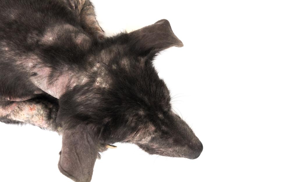
Another one of the most common issues brought to us here at My Pet Nutritionist is Atopic Dermatitis. Most commonly seen in dogs, it can also occur in the cat. Atopic dermatitis is a multifactorial disease process, but it is defined as a genetically predisposed inflammatory and pruritic allergic skin disease often associated with IgE (immunoglobulin) against environmental allergies. The prevalence of AD is thought to be 10-15% of the canine population. Symptoms will include: Scratching Chewing Licking Recurrent skin, ear and anal gland inflammation and infections Hair loss Thickening of the skin It is thought that genetic abnormalities, along with an altered immune system and skin barrier defects all play a role. So, lets take a look at AD in a little more detail. Where it comes from and what, if anything, we can do about it. Genes Multiple gene expressions involved in skin barrier function and inflammation have been seen to go awry in canine atopic dermatitis. They have been both up and down regulated. Gene expression is like the instruction pamphlet that comes with your washing machine. Issues in gene expression is like you being sent an instruction manual in a different language and inadvertently using the 60-degree wash on your cold wash items! It’s not going to end well. Well, cheeky genes who aren’t doing what they’re supposed to, often doesn’t end well either. In dogs suffering with atopic dermatitis, data has shown: 361 genes that instruct inflammation, wound healing or immune response were up regulated. 226 genes that affect skin barrier function showed decreased mRNA. mRNA is a messenger; so low concentrations result in fewer messages. Findings here This is why certain breeds are known to suffer with AD, specifically, German Shepherds, Labradors, Boxers, West Highland White Terriers, French Bulldogs, Bull Terrier, Lhasa Apso, Springer Spaniel, Poodle, Dachshund, Miniature Schnauzer and Pugs. That said, these predispositions are also linked to geographical area, which also demonstrates how the environment can interact with genes. There is much attention being paid to the idea of epigenetics; how behaviour and the environment can affect how genes work. This is why, even if your dog is genetically predisposed, you shouldn’t throw in the towel, just yet. Skin Barrier Defects The skin is the first line of defence in our trusty immune system. This goes for our dogs too. If the skin isn’t as robust as it needs to be, then pesky allergens or irritants can get in and start to knock on doors of our second line of defence; inflammation. It is believed that part of the defects found in the skin barrier in AD cases is due to decreased concentrations of filaggrin. Not only can this be due to a genetic encoding error, but certain enzymes in the skin can breakdown filaggrin. The more enzymes, the more filaggrin gets broken down. Filaggrin is a protein found in the skin, and it binds to keratin filaments. This forms a structure within the skin cells. Filaggrin is essential to skill cell maturation, and in forming the outermost protective layer. It also forms part of the natural moisturising substance found in the skin; without it,cells dry out. This is why low levels of filaggrin result in dry, cracked skin. There is also evidence that filaggrin associated atopic eczema is more likely to lead to food sensitisation and inhalant allergens. Findings here Despite there being a genetic cause for filaggrin deficiency, the importance of maintaining a robust skin barrier is essential. Please take a look at our top tips for promoting skill health here. Altered Immune Response As we have mentioned, if pesky allergens or irritants get past the first line of defence, they soon feel the full wrath of the immune response. What starts with inflammation, will venture to a targeted attack in order to eradicate the invader. In the case of a defective skin barrier, the system will regularly be exposed to potential threats, of which the immune system must deal with. This constant state of firefighting ensures a steady level of inflammation throughout the body, which is why cases of dermatitis are often paired with food sensitivities. This is why tackling chronic inflammation is a key part of a dermatitis plan. As you have likely gathered, ensuring a robust skin barrier is essential in managing atopic dermatitis, but so is modulating the immune response and inflammation. Here are our top tips in tackling those three areas. Polyphenols Polyphenols are known to interfere with pro-inflammatory mechanisms. They have been seen to act upon keratinocytes which attenuate skin inflammation. Not only that but they have been seen to hamper toxicity of bacteria which regularly colonises the skin of atopic dermatitis affected patients. Polyphenols have been seen to inhibit the activation, proliferation, and function of Th2 cells which are key players in allergic reactions. Specifically, in the inflammatory response in allergic reactions. This has also been demonstrated during re-exposure to the allergen, suggesting that offending allergens may even be reintroduced at a later stage. Polyphenols are micronutrients found in certain plant foods; they are packed full of antioxidants. Their original role in the plant is to protect against UV radiation and of course aggression by pathogens. For that reason, they tend to contribute bitterness, astringency, colour, flavour, odour and oxidative stability. Under the umbrella of polyphenols, you will find phenolic acids, flavonoids, stilbenes and lignans (and a few more). Along with being anti-inflammatory and anti-microbial, the antioxidant capacity of phenolic compounds is also key to skin health. Oxidative stress is the imbalance between free radicals (the exhaust fumes of work) and antioxidants. Oxidative stress can damage the structure of the skin and negatively influence immune system function. Interesting, polyphenols have been seen to alter certain gene expression in the dog. Findings here So, all things considered, they are pretty nifty things to offer in your dog’s fresh food diet. Phenolic Compounds for The Dog Blackberries Raspberries Strawberries Blueberries Apple Pear Curly Kale Broccoli Green Tea (organic and decaffeinated) Parsley Chamomile Pomegranate Be mindful
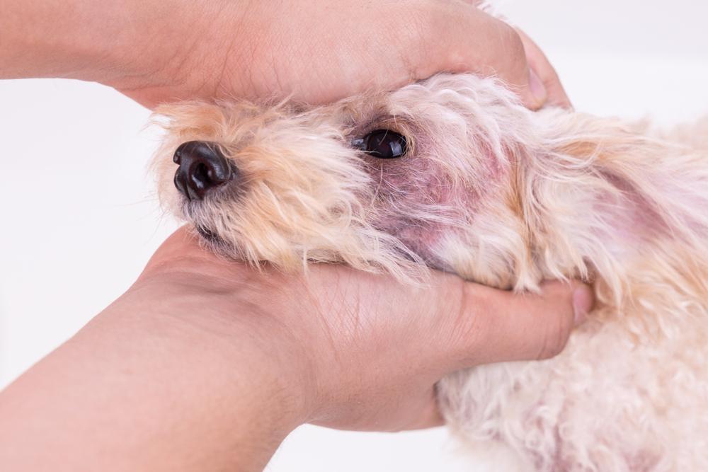
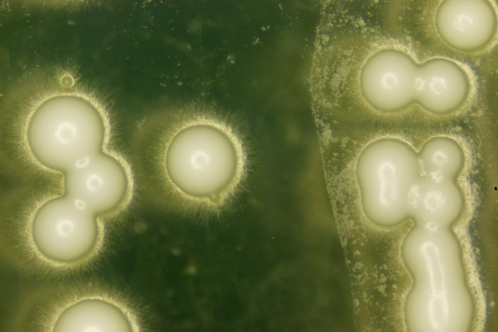
Another particularly common issue we see here at My Pet Nutritionist is yeast! More technically known as candida, it is actually harmless when kept in check. The issue is when our dog’s system goes a little awry and can’t keep those little blighters from colonising. Whilst it sounds like a military operation, the dog’s system functions very much like that. Let’s take a look at yeast in a little more detail and how we can armour dog’s defences for a fighting chance! What is Yeast? Candida is an opportunistic fungal pathogen. It is a normal part of the gastrointestinal flora and genital tracts. Healthy systems are more than capable of keeping it in check with their beneficial bacteria. The issue arises when there aren’t enough good guys to keep the bad guys in check. The good bugs we find in our dog’s gastrointestinal tract will compete with the bad bugs for food sources and attachment sites. As we know there are a number of factors that can skew the microbial community found in our dog’s system. Antibiotics Broad spectrum antibiotics target all bugs,so whilst they get rid of the ones running amok, they also wipe out the beneficial ones too. Whilst the microbiome can recover to some extent, eventually; this takes time and there’s evidence which suggests key species are never fully repopulated. Findings here Stress There are clear links that stress, whether physical or emotional, affects the composition of the gut. On many occasions, stressor infliction significantly reduces the good guys. This has been associated with increased inflammation in the gut, which then continues the cycle. Findings here The Environment There is a huge amount of data which shows exposure to environmental pollutants significantly alters the gut microbiome. This includes bisphenols, phthalates, organic pollutants, heavy metals and pesticides. Bisphenol is an industrial chemical used to make hard, clear plastic. It is also found in epoxy resins which is often used as a protective later in some metal food and beverage cans/tins. Bisphenol has been linked to reduced microbiome diversity, and a significant decrease in protective bacteria. Phthalates are plasticisers and stabilisers found in vinyl flooring, clothing, detergents, personal care products, toys, medical equipment, and plastic packaging. Because they are non covalently bound to materials, they can leach into the environment. Phthalate exposure induces microbiota changes and has been known to inhibit the synthesis of certain short-chain-fatty acids. Heavy metals are associated with reduced microbiota diversity along with the altered metabolism of vitamin E and bile acid. Findings here Pesticides are renowned for altering the gut microbiome. This not only includes the pesticides administered to pets (flea and tick treatments), but also those found in the environment, particularly those sprayed on public footpaths. What is also worth considering is that antibiotic use has been seen to increase bioavailability of pesticides within the body. Findings here You are what eat! Of course, nutrition also plays a part in modulating the gut microbiome. But we always knew that. We are all pretty selfish in evolutionary terms, and this includes the bugs we find throughout our body. Our ultimate aim, and theirs, is to survive. So, we just need to modify how many of them actually do. This means keeping the colonies of good bacteria strong, so offering our dogs a diet full of pre and probiotic foods! Prebiotics are like the fertilisers in the garden, they help to feed and grow the beneficial bacteria in our garden. Probiotics however contain live organisms which can contribute to the population of the garden. Findings here There are a number of prebiotic foods suitable for dogs and they include mushrooms, chicory root, garlic and dandelion greens. Probiotics include fermented food, but supplements are available; soil-based are a good call for your canine friend (hold off on the fermented food until later, if your dog has already developed an overgrowth). Yeasts seem to like sugar as fuel, so diets high in grains, starches and other carbohydrates seem to contribute to an overgrowth. Generally, certain beneficial bacteria will metabolise these sugars, keeping candida in check by disrupting its food supply, but in the absence of good bacteria, candida is partying it down at the all-you-can-eat buffet! Immunity A weakened immune system is also a huge risk factor for developing a candida overgrowth. Whether this has been a natural progression over a period of time for a range of factors, or even due to medications like steroids. Immune function has natural peaks and troughs, young dogs and ageing dogs naturally have a lower function, but pre-existing conditions along with a range of medications can affect it too. The immune system is like a nosey neighbour. It keeps tabs on everything going on in the body and knows when something isn’t quite right. When it identifies something as non-self, it sends the army to fight the foreign invader and, all being well, wins, before sitting back down with its cup of tea. The issue arises when it can’t get up from its chair and so the foreign bodies are left to invade. There are a number of factors which can compromise immune function from sleep, to stress and nutritional status; there are several key nutrients essential to its performance. So, supporting this is essential in the prevention and treatment of any bacterial overgrowth. Top Tips: Adequate rest Reduced exposure to stress Nutrient dense diet withVitamins A, C, D and B’s, along with Zinc, Selenium, and Iron Address any pre-existing issues like inflammation in the digestive system or poor pancreas function which can contribute to poor nutritional status Findings here Careful use of medications like antibiotics, NSAIDs and steroids Findings here on Vitamin D Findings here on Zinc What if your dog has already got an overgrowth? If indeed your dog has developed a candida overgrowth, you will notice symptoms like: Ear infections Greasy coat Sores Odour (yes, that cheesy type of smell) Green/yellow discharge Crusty/flaky skin Hair loss Itching/scratching Incessant licking/grooming of an area due to irritation Rust
Here at My Pet Nutritionist, skin issues are one of the most common complaints from dog owners. Whilst there are a number of factors that can result in not-so-comfortable skin for your companion, there are also some top tips that can start you off the right foot. So, let’s take a look at the skin in a little more detail and what can do to promote its health. The skin is the largest organ of your dog’s body. It consists of three major layers: The Epidermis – (Epi – upon or above) this is the outer layer of skin, the protective layer. The Dermis – the dermis supports and nourishes the outer layer. It provides strength and elasticity. Here you will find collagen fibres, sweat glands, sebaceous glands, and hair follicles. You will also find cells that release histamine and other inflammatory mediators when faced with an allergy or injury. The Subcutis – (sub meaning under or below) this in the innermost layer of the skin, where you will find fat and muscles. Subcutaneous fat provides insulation, padding and storage for reserve energy. Not only does the structure of the skin prevent water and electrolyte loss to help maintain body homeostasis, but it forms a protective barrier which helps protect against infections, parasites, and the elements. This is the often-forgotten role of the skin –that it forms part of the immune system. We have three lines of defence in the immune system: The first line of defence are the physical barriers, the skin and mucous membranes of the gut and respiratory tract. The second line of defence is defined as innate immunity. This system surveys and neutralises pathogens by mounting an inflammatory response. This system communicates with the third line of defence which is adaptive immunity. Adaptive immunity provides a specific and tailored response, deploying T and B cells. Ultimately, for us, or our dogs to survive and thrive, we, and they need robust immune defences, so it makes sense to want to strengthen our first line as much as possible. 1) Sleep! In the dermis of your dog’s skin, you will find connective tissue which contains collagen. Collagen fibres play a vital role in maintaining structural integrity and it is supported by hyaluronic acid (in ageing human, skin hyaluronic acid is often absent leading to the presence of fine lines and wrinkles). Cortisol, however, significantly decreases the synthesis of hyaluronic acid. Findings here As we know, cortisol is one of the main stress hormones, released in times of fight or flight. In short, stress results in lower hyaluronic acid, impacting collagen and therefore skin structure. There are many stress triggers for your canine companion, but lack of sleep is often missed. Not only does low sleep duration influence the perception of stress for days following, but sleep deprivation is in fact a form of neurobiological and physiological stress (or torture, for us at My Pet Nutritionist). In 1894, Marie De Manaceine was fascinated with sleep deprivation. She had established that mental disturbance resulted from partial insomnia, but she wanted to know more. So, in her Lab, she recruited puppies. 10 to be specific; aged 2, 3 or 4 months old. Whilst they continued to be fed by their mother, she kept them in constant activity. In short, she deprived them totally of sleep. After 96-120 hours, the puppies were irreparably lost. We shed a tear when we read this! What is interesting (despite it being macabre), is that when puppies were starved, they could be saved after 20-25days. This wasn’t possible when they were sleep deprived. Marie found that sleep deprivation significantly affected the puppy’s brains. When they were starved, the brain was left almost spared. But, in the absence of sleep; fat degeneration, blood vessel abnormalities and haemorrhaging occurred. Findings here In a world that never sleeps, both us and our dogs really need to. Adult dogs, in a laboratory setting, when left, will sleep on average for around 13 hours per day. Puppies can sleep anywhere between 18-20 hours per day. Dogs are diurnal, meaning they are active during daylight hours. Rest occurs during dark periods with activity increasing the two hours before light. Dogs have a natural rest period around noon and then reduced activity during the afternoon. Findings here Interestingly, the experiences that your dog has, can affect the type and quality of sleep they experience. Studies have found that after a negative experience, dogs will fall asleep more quickly. It is thought that this is a protective sleep, in response to stress. This should be in the forefront of your mind. It is all too easy to attribute an “exhausted” dog to the busyness of the day. Be mindful, their sleeping habits could be more indicative of their experience. Findings here Sleep is vital for your dog’s overall health, but also in helping their body synthesise the compounds it needs to build a robust skin. Build in plenty of rest time for your dog, at the times they would naturally do so. 2) Feed Fresh Food! We probably sound like a stuck record here at My Pet Nutritionist, but fresh really is best! Processed, high glycaemic index foods wreak havoc with skin! There are a few different reasons, but glycation is one of them. Glycation is the modification of proteins or lipids after exposure to sugars. You may notice them referenced as AGEs, advanced glycation end products. Glycation leads to a loss of protein function and impaired elasticity of tissues like blood vessels, tendons and of course skin. AGEs have been associated with many metabolic disorders and are closely related with renal failure and diabetes. They also increase with age. AGEs can be synthesised in the body, but total load also includes dietary exposure. It is clear that foods ranking high on the glycaemic index result in a higher accumulation of AGEs. Not only that, but maillard reactions, those generated in the heat processing of foods, also result in AGEs. Another reason why