Haematomas are one of the more common soft tissue issues we tend to see here at My Pet Nutritionist. The most frequently occurring Haematoma is the Aural Haematoma; a subdermal haematoma found on the ear, however these benign masses can also be found on the spine, the brain, the heart, or anywhere else in the body. This blog will look at some of the types of haematoma, symptoms, diagnoses, and how we can offer support when a dog has a haematoma. What is a Haematoma? Similar to a bad bruise, a haematoma is a collection of blood which has formed in an organ or body tissue, typically as a result of a ruptured blood vessel. Haematomas are often mistaken for a tumour, due to their tumour-like appearance. Haematomas present as hard, raised lumps, which are usually very painful, whereas a bruise is a flat, malleable discolouration of the skin which may be a little sore to the touch. Where bruises usually heal with no additional medical help, haematomas sometimes require surgical intervention, such as drainage as they can become so large, they can cause life threatening issues due to the inability to get sufficient blood flow to a certain area, or they may become infected. Types of Haematoma Let’s discuss a few of the more common haematomas, their symptoms, and causes. Aural Haematoma As previously mentioned, haematomas of the ear are the type we see most often. These usually present on the flap of the ear (scientifically known as the ‘pinna’). When the blood vessel in the ear is damaged, blood leaks into the area between the skin and cartilage of the ear, which causes a pool of blood to form. Depending on the severity of the bleed, the ear position may change as the pool of blood may interfere with the muscles in the ear, as well as the ear canal, due to swelling. Aural Haematomas are extremely painful to the touch, so avoid touching the ear too much so as not to cause pain and distress in your pet. Aural Haematomas happen when there’s trauma to the ear, usually caused by dogs shaking their heads during ear infections and ear mite infestations, or by excessive itching of the area in allergy dogs, or dogs with insect bites on the neck or head areas. Aural Haematomas are also fairly common in dogs who have been a victim of a dog attack when the dog bite hasn’t broken the skin. Studies suggest that Aural Haematomas are not as a result of autoimmunity, but could be caused by other immunological events, such as infection control in the presence of a pathogen, or histamine responses in the presence of an allergen. Findings Here Aural Haematomas can heal on their own, however they often result in ‘cauliflower ear’ – the pinna appears crumpled/intricately folded. The science behind this malformation of the ear following a haematoma, is down to the substance released during the trauma of the area. The fluid which leaks between the cartilage and the skin, is a fibrotic rich serum. When the haematoma heals, the body reabsorbs the leaked fluid, which leads to fibrosis in the pinna. When fibrosis occurs, the fibrotic tissues in the area contract, which causes the folds often seen. Findings Here Haematomas on the Brain Haematomas can be found on the brain, and can be Subdural or Extradural Haematomas. Subdural Haematomas are often associated with underlying brain injuries, whereas Extradural haematomas are usually down to skull fractures, and damage to a meningeal artery (the main blood supply for the brain). These are generally the most dangerous type, and have a variety of symptoms, including vomiting, lethargy, paralysis on one side of the body, confusion, and uneven pupil sizes. In cases where the haematoma is located on the brainstem, upper (cervical) spinal pain is common too. Haematomas on the brain are most commonly seen following trauma to the head from vehicle accidents. Findings Here Findings Here Findings Here Findings Here Studies show Cerebral Haematomas can also be caused by an infestation of French Heartworm (Angiostrongylus vasorum). This is just one of the ways the parasite causes major neurological problems. Findings Here While the prognosis for those diagnosed with any subdural haematoma may be a scary one, there is evidence to suggest a veterinary treatment plan is available, and successful for the condition. A study carried out on a large breed dog, kicked in the head by a horse, shows a full recovery within 15 months of intercranial surgery, antimicrobial therapy, and corticosteroids. Findings Here Spinal Haematomas Spinal Haematomas can be present for a number of reasons, and can be debilitating during recovery. Intervertebral Disc Disease (IVDD) patients may suffer from a spinal extradural haematoma, as a result of trauma during the herniation of the affected vertebrae. Spinal compression caused by Pneumorrhachis (air bubbles stuck in the spine) can also lead to haematoma formation. Findings Here Findings Here Findings Here There is also evidence to suggest that bites from venomous snakes may also contribute to haematoma formation on the spine. The other most common cause of spinal haematoma is trauma during spinal surgery. Findings Here Findings Here Tracheal Haematomas Haematomas in the trachea can cause acute (sudden) respiratory difficulties/blockages, due to the airway being blocked by the raised haematoma. These are most commonly discovered following surgery, and are caused by the intubation tube used. When the wrong type, or size of intubation tube is used, or the tube is not handled carefully, trauma occurs in the trachea which can cause bruising, or damage to the blood vessels, resulting in the formation of haematomas. Trauma following neck surgery may also contribute to the formation of Tracheal Haematomas, but there are studies to suggest this is the case, and others to suggest it isn’t the case so more research needs to be carried out to determine a definitive answer. Findings Here Findings Here Findings Here As with other types of haematoma, trauma from non-surgical events can also cause haematomas. A
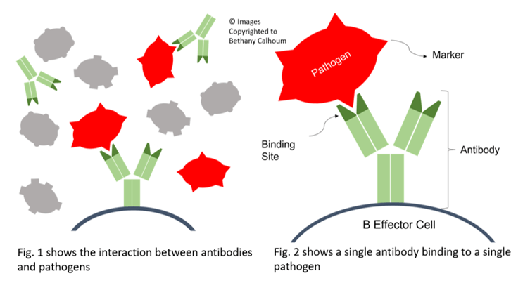
In Part 1, we discussed what Leaky Gut is, what autoimmunity is, and possible causes and symptoms of both. Read on to find out how they are linked, and more about the autoimmune diseases most commonly affected by Leaky Gut, as well as how we can support the body through diet, and supplements. How are Leaky Gut and Autoimmunity linked? A healthy gut microbiome is incredibly important as around 90% of the immune system is located in the gut! That’s quite a staggering figure, but it shows the importance of keeping the gut healthy and the microbiome strong. Let’s take a deeper look at some of the most common autoimmune diseases, and their link with Leaky Gut. Thyroid Issues One of the main issues with Leaky Gut and Autoimmunity, involves the thyroid. The body attacks the thyroid tissue as it recognises it as a foreign body. The reason the body sees thyroid tissue as a threat, is down to molecular mimicry. When the immune system releases antibodies to get rid of a threat, they bind at what is known as the ‘active site’, or ‘antigen binding site’. Antibodies are Y shaped proteins, and on the tips of the Y, the binding sites are found. These are a specific shape, to match the proteins on the antigens (the threatening particles). Take a look at the diagram at the top of this blog! Despite food particles clearly being very different to thyroid tissue cells, some of the attached proteins are the same shape on both the food particle and on the thyroid tissue cells. Gluten and Casein (dairy) are particularly alike to thyroid tissue cells, so when an antibody detects the protein it fits with, and binds to it, there’s a high chance it may be thyroid tissue instead of its real target; the food particle. Findings Here Findings Here Inflammatory Bowel Disease (IBD) A condition we hear a lot about, particularly on a professional basis as well as on social media posts when people ask advice on their poorly pets, is Inflammatory Bowel Disease. As per it’s name, this involves inflammation in the bowel, which can be as a result of Leaky Gut. When looking at IBD, diet is so important – many processed foods contain emulsifiers, which can include Cellulose Gum, and Polysorbate-80 (though this one is most inhuman foods, not pet foods). These have been found to interrupt interactions between the bacteria in the intestine, and the gut wall; resulting in the gut wall being less protected than it would be without the presence of these substances. This lack of positive interaction, teamed with the already permeable gut wall (due to Leaky Gut) can cause the onset of IBD. Findings Here Small Intestine Bacterial Overgrowth (SIBO), Yeast, and Candida can also contribute to IBD (and are all symptoms of Leaky Gut), which causes inflammation of the bowel, which further increases the risk of IBD onset. Studies show a huge affect on gut permeability when up-regulation of the protein called Zonulin is present. Zonulin helps regulate the permeability of the small intestine, but is detrimental in high numbers as it causes the gut to become more permeable. It is secreted by numerous organs within the body, and can be linked to Leaky Gut and the onset of IBD. Findings Here Findings Here Findings Here Immune-Mediated Haemolytic Anaemia (IMHA) IMHA is one of the more worrying autoimmune diseases, with a mortality rate close to 70%. There are many reasons a dog may be diagnosed with IMHA, including Vaccine Induced IMHA. When a dog has IMHA, the body is attacking it’s own red blood cells, which are important for transportation of oxygen from the lungs to all parts of the body for healthy muscle usage. IMHA can be caused in some rare cases, by a deficiency in Vitamin B12, which can be linked back to Leaky Gut. IMHA is also often as a knock on effect of other autoimmune diseases like Systemic Lupus Erythematosus. Findings Here Findings Here Diabetes Largely linked to Leaky Gut, Diabetes mellitus often requires lifelong medication. Similarly to the mimicry of thyroid tissues by antigens like Gluten and Casein, the onset of diabetes can be facilitated through normal cells being attacked incorrectly by the antibodies circulating the bloodstream. With diabetes cases, the immune reactions affect, and damage pancreatic beta cells (responsible for producing and secreting insulin), which then causes the over-production of cytokines, which in turn causes insulin resistance within the body. Healing the gut, and decreasing gut permeability may help relieve sufferers of diabetes symptoms. Studies show Type 1 Diabetes can be caused as a result of gut damage, but can also cause gut damage. Findings Here Findings Here Findings Here Findings Here Immune-Mediated Trombocytopenia (ITP) ITP is a platelet disorder, in which sufferers are unable to properly clot blood due to low platelet counts. Pathogenesis of ITP as a result of leaky gut has been proven to be due to imbalances in the gut microbiota, and the presence of cytokines which interfere with metabolism of fats. Patients with Leaky Gut, as we know, have a very imbalanced microbiome as bacteria leaks out through the channels in the gut wall. Certain strains of bacteria play an essential role at keeping ITP at bay, but are found to be of low levels in those diagnosed with ITP. When these helpful bacteria are leaked from the gut, cytokine production is increased, which then affects the metabolism of fats, which in turn causes pathogenesis of ITP because the lack of fat metabolism causes a lack of available fatty acids to enable the blood to clot. Findings Here Findings Here Findings Here Rheumatoid Arthritis The previously mentioned protein called Zonulin plays a part in Rheumatoid Arthritis (RA); a joint related autoimmune condition. Just like in IBD, when Zonulin is over-represented, the gut permeability cannot be controlled, and allows useful and harmful substances to enter the blood stream, which are then detected as threats by the immune system. The upregulation of
At My Pet Nutritionist, we regularly help people with their pets suffering with Leaky Gut Syndrome. There is a lot of evidence to link Leaky Gut with a variety of autoimmune diseases; issues we so very often offer support for. Read on to learn more about Leaky Gut, and the role it plays in autoimmunity. What is Leaky Gut? The condition is growing more and more common in both us humans, and our canine counterparts, and can lead to some pretty serious health issues, long term. The name ‘Leaky Gut’ does somewhat give the game away, but let’s look deeper into what actually happens in the gut of a normal dog,compared to one with Leaky Gut. In healthy individuals, after eating, the food passes through the gut. The gut consists of the stomach followed by the small intestine, followed by the large intestine (known as the ‘colon’), through which nutrients from the digested food are absorbed, before the waste is pushed out through the rectum, then anus. To enable a large surface area, for optimum nutrient absorption, the small intestine is lined with small finger-like structures called villi, which themselves are covered in even smaller finger-like structures, known as microvilli. The gut also houses lots of good bacteria to aid digestion – the colonies of good bacteria, along with yeast cells, any viral particles, or parasitic burdens, are collectively known as the ‘microbiome’. The gut wall is extremely thin, to allow efficient nutrient absorption. The cells lining the gut stay close together, and are supported by the interactions of immune cells, and good bacteria in the gut. In those suffering with Leaky Gut, inflammation occurs in the gut for various reasons, which causes the tight intestine wall to permeate, creating microscopic channels between the cells. Proteins/partially undigested foods then leak out through these channels and are detected by the immune system as a threat, causing a histamine response to occur, which is why one of the most common symptoms of Leaky Gut, is food intolerances. Other symptoms of Leaky Gut include: – Autoimmune Diseases – Issues with stools or sickness – Joint issues – Yeast – Problems concerning other major organs in the body – Hypothyroidism – Changes in behaviour; often anxious behaviour and short tempered behaviour Possible Causes of Leaky Gut include: – Over-use of vaccines; the adjuvants damage the gut flora – Use of certain pharmaceuticals ^ Flea, tick, and worm medications; they disrupt the gut microbiome by not only eradicating the visiting parasites (or often lack thereof), but the good bacteria too. ^ Antibiotics; these wipe out the good and bad bacteria ^ Antihistamines; these can interfere with the production of mucus in the gut, and can also interfere with the proper functioning of Diamine Oxidase (DAO), which is the enzyme responsible for breaking down, and removing histamine from within the gut. ^ NSAIDs and Steroids; these can cause ulcerations in the gut and interfere with mucosal production. – SIBO and Yeast overgrowth; Small Intestine Bacterial Overgrowth and Yeast damage the gut lining – Diet; feeding a dry food diet can put stress on the gut. Kibble often contains Glyphosate, which is an antibiotic herbicide and is toxic, as well as very damaging to the gut. Diets inclusive of legumes and other high-lectin content pulses, nightshades and vegetables may contribute to Leaky Gut as lectin causes poor gut integrity. Microscopic moulds often found on kibbles, known as mycotoxins are also detrimental to gut health, contributing to Leaky Gut. – Stress can have a huge effect on the gut integrity, as stress leads to inflammation – Ageing; as our dogs age, the microbiome becomes less diverse which leads to gut damage. Findings Here Findings Here Findings Here Findings Here Read our full Gut Dysbiosis blog here What is Autoimmunity? To better understand the link between Leaky Gut and Autoimmunity, we need to understand more about autoimmunity, what it is, and how it affects our pets (and us too!). Autoimmunity is sadly fairly common in both humans and pets and is often overlooked. When an individual has an autoimmune disease, the immune system releases antibodies and T-Killer Cells (cells of the immune system which target and kill cells infected with viruses and cancers) even when they are not in the presence of a necessary target, which causes them to attack normal, previously healthy parts of the body. In layman’s terms, the body attacks itself! There are more than 80 different autoimmune diseases in existence, though not all of these affect dogs. Some of the most common autoimmune diseases we see in dogs are: – Hypothyroidism – Inflammatory Bowel Disease (IBD) – Immune-Mediated Haemolytic Anaemia (IMHA) – Diabetes – Immune-Mediated Trombocytopenia (ITP) – Rheumatoid Arthritis – Hypoadrenocorticism (Addison’s Disease) – Periodontal Disease – Degenerative Myelopathy – Immune-Mediated Polyarthritis (IMPA) – Systemic Lupus Erythematosus (SLE) – Discoid Lupus Erythematosus (DLE) Symptoms of Autoimmunity These will, of course, vary depending on which autoimmune disease an individual has, but general signs and symptoms of autoimmunity, which may spark a look into further investigation with your veterinarian, include: – Constant lethargy – Racing, or very slow heartbeat – Weight loss (often dramatic) – Increased panting – Collapse – Excessive drinking and urination – Seizure activity – Discolouration of gums and skin – occasionally yellowish as a result of jaundice. – Hair loss or coat texture changes – Regular vomiting and diarrhoea – Increased temperature – Behavioural changes, including aggression or depression Findings Here Read Part 2 here to find out more on the autoimmune diseases affected by Leaky Gut, and how we can support the body, naturally. If you feel your dog may be experiencing Leaky Gut, or an autoimmune disease, seek veterinary attention, and book in for a consultation with one of our team! Team MPN x
Being a fairly common health complaint in dogs, particularly larger breeds of dog, here at My Pet Nutritionist we feel it’s important to understand what Laryngeal Paralysis is, what it looks like, what causes it, and how to support the body. We will discuss all these points in this blog! What is Laryngeal Paralysis? Laryngeal Paralysis is a disease which involves the Larynx; commonly known as the ‘voice box’. The larynx is a box-like structure which connects the throat to the windpipe (trachea); and is comprised of various plates of cartilage known as ‘Arytenoid Cartilages’, housing the vocal cords. As well as enabling vocalization in all mammalian species, the Larynx closes off the top of the trachea to ensure food and water are not inhaled. When an animal takes a deep breath, the larynx opens wider to allow for more air to be taken in. The larynx is surrounded by muscles called ‘Laryngeal muscles’ which help keep it stable. As with all muscles, if the nerves inside become damaged, it causes the muscle to relax. If the laryngeal muscles become weakened or paralysed due to nerve damage, the cartilage of the larynx will collapse inwards, as the cartilage is no longer stabilised by the muscles. When the muscles are weak or paralysed and the larynx collapses, this is called Laryngeal Paralysis. Laryngeal paralysis can be congenital (present at birth), hereditary (passed on genetically through generations) or acquired (due to trauma or as a knock-on effect from other health conditions). Like many conditions, some breeds are at a higher risk of developing Laryngeal Paralysis than others. Generally speaking, this disease affects larger breeds of dog. Most commonly affected, is the Labrador Retriever. Different breeds are more commonly affected by different types of Laryngeal Paralysis. Breeds most at risk of acquired Laryngeal Paralysis, usually in middle aged to older dogs: Labrador Retriever Great Dane Irish Setter Newfoundland St. Bernard Breeds most at risk of hereditary and congenital Laryngeal Paralysis: Leonberger Bouvier des Flandres Siberian Husky Bulldogs (various types) Studies also show a higher risk of developing Laryngeal Paralysis for neutered male dogs over entire males, or entire/neutered females. Findings Here Findings Here Findings Here Symptoms There are many symptoms for Laryngeal Paralysis; let’s take a look! Excessive/Noisy Panting Dogs with the condition will likely pant more than is normal for that dog, especially during humid weather and when stressed or after exercise, and this panting is often quite noisy. Lethargy They may become lethargic or wish not to exercise as a result of Laryngeal Paralysis. Change in Bark Many owners notice a change in the dog’s bark; just like in humans when one’s voice may change, a dog’s bark also has the capability to change if they have a collapsed larynx. Choking, Coughing or Gagging When eating or taking a drink, the dog may choke, cough or gag – this is due to the windpipe not being fully shut off from the throat, and the width of the larynx being extremely narrow. Coughing may mechanically force the larynx to open and allow food and water to enter. As drinking and eating becomes more difficult, those suffering with Laryngeal Paralysis are also more susceptible to Aspiration Pneumonia. Behavioural Anxiety You may notice an increase in behavioural anxiety due to the feeling of vulnerability, as well as respiratory distress due to the narrow opening of the collapsed larynx. Dehydration As water intake becomes more difficult for those suffering with the disease due to the narrow opening, the dog may become dehydrated. Gums will become greyish, dark red or purple due to lack of proper blood circulation as a result of dehydration. The gums also become tacky when the dog is dehydrated. Difficulty Thermoregulating Dogs with Laryngeal Paralysis are more susceptible to heatstroke, even in mildly warm temperatures, is another symptom of Laryngeal Paralysis, and can result in collapse. If your pet is showing signs of heatstroke (vomiting, shaking, seizures, lethargy, panting, glassy eyes, agitated whining, drooling, accelerated heart rate, unconsciousness) it’s imperative to seek veterinary care immediately (though don’t put your dog in a hot car!). Your dog may display multiple of the above symptoms of varying degree. Diagnosis So, how would the vet diagnose Laryngeal Paralysis? There are a few routes to diagnosis of Laryngeal Paralysis, but all will start off by looking at the medical history of the dog, and clinical presentations. Some vets may run X-rays of the chest to rule out problems within the chest cavity, and run blood panels, and urinalysis to rule out infection before examination of the Larynx itself. To avoid sedation, there is evidence to suggest that a suitable method of formal diagnostic testing for Laryngeal Paralysis is by performing an echolaryngography, through the use of ultrasound. Large dogs can be tested on the floor or table, while smaller breeds can happily reside on the lap of the sonographer to reduce risk of false results due to stress. Echolaryngography is a safe, and effective way to diagnose Laryngeal Paralysis. Findings Here Findings Here Another common method, used to diagnose lightly sedated dogs in order to reduce risk of false results due to full anaesthesia (which may cause the laryngeal muscles to relax), is through a transnasal laryngoscopy, where a video endoscope tube is inserted through the nostrils and down the throat to have a good visual of the larynx working. Studies prove this method to be as accurate as a traditional laryngoscopy, whereby the patient may require heavier sedation due to potential gag reflexes following intubation by mouth. Findings Here Findings Here Findings Here Causes Trauma Trauma to the neck area is often a cause of Laryngeal Paralysis. This can be through repeated use of unsuitable training tools which constrict around the neck, poorly fitting flat collars on dogs who pull, or even through freak accidents involving the neck area such as dog bites and subsequent deep wounds. We see many dogs who sadly develop Laryngeal Paralysis following a general anaesthetic; likely due
A common issue presented among our clients, readers, and followers here at My Pet Nutritionist is low stomach acid (hypochlorhydria).There are numerous signs and symptoms that your pet may have low stomach acid; let’s discuss the most common ones! Acid Reflux Low stomach acid will usually result in Acid Reflux, or it’s most severe form, Gastroesophageal Reflux Disease (GERD). When a dog has GERD, there is a backflow of stomach acid. Dogs presenting with the below clinical signs, are often prescribed Proton Pump Inhibiting drugs (PPI’s), which suppress acid secretion in the stomach, and can provide temporary relief, but can cause more implications in the longer term, and will not erase the cause of the low stomach acid, or its symptoms. Most commonly seen in brachycephalic dogs and cats, and those with shorter a oesophagus, low stomach acid is being seen more and more often in the animal health and nutrition industry. Read our Guide to Acid Reflux here Regurgitation Probably the most common sign of low stomach acid is regurgitation (bringing up partly digested food), bringing up bile (clear, yellow substance produced in the liver, and stored in the gallbladder for use during digestion), and sometimes bloody vomit. These various nauseating problems occur due to the mixture of digesting/partly undigested food, known as chyme, sitting in the stomach for prolonged periods of time. When it sits in the stomach for so long, inflammation worsens in the area, which ultimately causes food or bile to come back up, since it is unable to go down! A popular supplement used as part of many dogs’ battles against regurgitation, is Deglycyrrhizinated Liquorice (DGL)which is a great anti-inflammatory for the stomach, and helps heal the gut. Findings Here Indigestion Dogs and cats fed a raw diet while suffering from low stomach acid, will often be unable to properly digest meat and bone. Pets with indigestion will often have pieces of bone and fresh-looking meat in their vomit. In order to digest bone and meat (or other sources of protein), the gastrointestinal juices must be acidic. If there is little acid available, the pet will be unable to digest and utilise the bone and meat. During fasting (between meals), the dogs gastrointestinal juices are around pH 7.3, which is almost neutral. In anticipation of food, during eating, and during the process of digestion, the gastric juices drop to a very acidic pH of 1 to 2 – that’s not too dissimilar to the pH level of battery acid! Findings Here We often recommend a gently cooked diet as per our balanced recipes for those suffering with low stomach acid, as cooked meat and bone substitutes are much easier to digest. Lip/Air Licking Another very common sign of low stomach acid is lip licking, often called ‘lip smacking’, as well as licking the air. These are not only signs of pain, but also signs of nausea in both dogs and cats. Drooling and lack of appetite are also signs of nausea in both species. Stomach Pain, Bloating and Gas Due to the inability to digest food properly and efficiently, stomach pain (particularly after eating) is common in those suffering with low stomach acid production. Dogs especially, often display unusual behaviours like bowing (just like the ‘downward dog’ yoga position!) when their stomach is sore. This position can help relieve the pain. Some pets will look sharply at their stomach when in pain, which is a very subtle sign, so often one missed. Cats will often become more vocal when in pain, or lose their appetite. Stomach pain can also be caused by bloating, and excessive gas. The inflammation of the stomach can lead to visible bloating. The gasses produced by the chyme mixture in the stomach will also further add to the bloating. In order to release these gasses, owners of pets suffering with low stomach acid will often notice more passing of wind through the anal passage, or through burping. Probiotics and gut healing protocols can help reduce these symptoms. Another cause of bloat and excessive gas is bacterial overgrowth in the gut. Known as SIBO (Small Intestine Bacterial Overgrowth), the overgrowth of bacteria alters not only gut motility, but also affects the gut-brain axis. Findings Here Findings Here Burping/Empty Swallowing Burping and empty swallowing (regular swallowing despite the pet not eating or drinking beforehand) are signs the pet has low stomach acid. Due to the low stomach acid content, the chyme will produce gasses which will cause burping and the feeling of sickness, hence the swallowing. Dogs and cats may also swallow when they have regurgitated, which would appear as empty swallowing. Constipation and Diarrhoea Irregular bowel movements and consistency of bowel movements can be a sign of low stomach acid in dogs and cats. Due to the lack of appropriate levels of stomach acid in these individuals, their metabolic rate decreases, meaning that important parts of the diet will take differing times of absorption, which can play havoc on the bowel. Pets who struggle with indigestion of raw bone may struggle with diarrhoea. A large part of the problem when it comes to irregular bowel issues, is down to bacterial overgrowth in the gut. As previously mentioned, SIBO has a huge impact on the gut-brain axis, as well as gut motility, leading to malabsorption of nutrients which can ultimately lead to both chronic and non-chronic diarrhoea and constipation. Conditions such as IBD may be diagnosed following SIBO in dogs with low stomach acid. Findings Here Bad breath As the chyme is stuck in the digestive system for such along time, it can lead to bad breath. Many pet owners associate bad breath with oral hygiene/dental issues, which is absolutely a possibility. These owners are often startled to see their pets have perfect oral health; yet are still suffering from bad breath. Digestive issues are a very common cause of bad breath, so it’s important to look into these too, especially SIBO. Focusing on the gut health should dramatically improve bad breath.
Iodine is an extremely important mineral to include in your dog’s diet. It has many uses in the body and is the backbone of many bodily functions. At My Pet Nutritionist, we understand the importance of micronutrients, and strive to educate pet owners in how to feed their pets with maximum health benefits. Let’s look at what roles iodine plays in the body, and where it can be found. The Role of Iodine in the Body Iodine is a trace mineral which cannot be made by the body. As it is so important, and cannot be produced by the body, it must be consumed in the diet of all mammals. Iodine is essential for healthy functioning of the thyroid. The hormones used by the thyroid, thyroxine and triiodothyronine, cannot be made without iodine. These hormones support a healthy metabolism, as well as protein creation and keeping enzymatic functions under control in the body. Linked to the support of enzymes, an iodine rich diet enables the body to use calorific intake as energy instead of being stored as fat. While doing this, iodine also helps rid the body of harmful heavy metals like Mercury. Iodine can also aid cancer prevention by causing a process called ‘Apoptosis’ to occur. Apoptosis is the self-destruction of cancerous cells. Acting as an antioxidant stimulator is another of iodine’s roles in the body, and in turn helps keep the immune system strong. If we compare canines to humans with regards to iodine turnover in the thyroid, the turnover of iodine in the canine is far more rapid than that in the human. Dogs are not as good at conserving iodine stores as humans are, and also deposit more in faecal waste, making iodine consumption extremely important. Findings Here Findings Here Sources of Iodine Iodine is found in the highest amounts in sea dwelling vegetation, and animal based proteins. The amount of iodine in different sea vegetable based supplements varies, so it’s important to choose one with a good amount of iodine, but not too high an amount. Findings Here Kelp Seaweed – dried – 6635mcg per 5g Wakame Seaweed – dried – 210mcg per 5g Nori Seaweed – dried – 116mcg per 5g Cod – cooked – 186mcg per 100g Oysters – cooked – 109mcg per 100g Eggs – cooked – 26mcg per egg Beef Liver – cooked – 16mcg per 100g Prawns – cooked – 15mcg per 100g Leafy greens – steamed – 8.8mcg per 100g Low Fat Fish Iodine Deficiencies There are numerous health issues caused by a lack of sufficient amounts of iodine in the diet. Let’s explore those. Hypothyroidism Hypothyroidism is caused by a deficiency of the thyroid hormones, which is caused by a deficiency of iodine in the diet. Iodine is a huge part of the synthesis of thyroxine and triiodothyronine, so with a lack of it, these hormones cannot be produced in the amounts required by the thyroid to avoid hypothyroidism. Symptoms of hypothyroidism include weight gain due to decreased metabolic rate, difficulty regulating body temperature/warming up, baldness/excess shedding, lethargy, and other skin related issues. One study also suggests a link between hypothyroidism and behavioural changes, but this is an avenue not fully explored yet. Some commercially prepared diets have been shown to lack the correct amount of iodine a dog needs, vs home cooked with and without suitable iodine supplements added, so no matter what you feed, if any of the symptoms above are present in your dog, you may wish to book a consultation with one of our team. Read more about Hypothyroidism here. Findings Here Findings Here Cancer Iodine deficiency seems to stimulate follicular cell-derived thyroid cancer. It may not be the base cause of the cancer itself (known as the ‘initiator’) but certainly stimulates carcinogenesis. Thyroxine therapy is often used in thyroid cancer cases, to suppress the cancer. Studies show carefully supplemented diets with sufficient iodine content result in less serious cases of thyroid cancers. Findings Here Findings Here Studies also show links between cancers of the breast and iodine deficiency. Treating canine mammary cancer with iodine alongside antineoplastic drugs is proven to be very effective. Findings Here Goiters Goiters are swellings of the thyroid gland, which present clinically as a lump in the throat. Often a direct result of an iodine deficiency, goiters can be rectified by increasing the amount of iodine in the diet. Goiters can also be caused by hypothyroidism, hyperthyroidism, or even thyroid disease, so its important to seek veterinary attention. Findings Here Can You Give Too Much Iodine? The short answer is … yes! There are numerous studies on dogs given too much iodine, and iodine toxicity is a condition to be mindful of. At My Pet Nutritionist, of course we recommend a well balanced fresh diet, whether that is raw or cooked, as per our recipes, however if you are feeding a commercially prepared dry food, be mindful that the food may either lack, have the correct amount of, or even exceed the required amount of iodine for your dog; we just don’t know! Hyperthyroidism is a risk when the dog has too much iodine in the diet. Symptoms of hyperthyroidism include weight loss due to a vast increase in metabolic rate, swelling of the neck/thyroid gland, excessive urination, excessive thirst, excessive defecation, and choking/vomiting. Read more about Hyperthyroidism here. Findings Here If you have any worries about your pet’s diet, or want to improve their diet, please don’t hesitate to book a consultation with one of our team! Team MPN x
As a we are well and truly into the festive season, our team here at My Pet Nutritionist thought we would have some foodie fun! Here’s our doggie (and kitty, for the animal based ones!) rendition of 12 Days of Christmas, as we take a look at the benefits of the My Pet Nutritionist ’12 Foods of Christmas’! On the first day of Christmas my owner gave to me….Turkey! Probably one of the first foods we think of when mentioning Christmas, turkey is a great option for your dog over the festive period (and any other period too)! Often used when first transitioning to a raw or fresh diet, turkey is easily digestible, and low in fat; around 4.7g per 100g with no skin. Turkey breast is the lowest fat part of the turkey. Adding turkey skin does add a little fat. Turkey is roughly 30% protein, so a great protein source! As turkey is low on the Glycaemic Index (GI), it helps reduce the amount of cholesterol often associated with major health implications, and replaces it with ‘good’ cholesterol. Low GI foods are also great for diabetes sufferers because they don’t cause a blood sugar spike, unlike those food items higher on the Glycaemic Index. Turkey is a great source of selenium, which has links to reducing the risk of certain cancers, including those affecting the bladder,stomach, prostate, and lung. Selenium protects cells from damage caused by free radicals. Findings Here Findings Here Choline is another essential nutrient in a dog’s diet, which is found in turkey. With benefits to cognitive function, cardiac and hepatic health, nervous system and proper functioning of muscles, choline is important to include in the diet. Another great source of Choline is krill oil. Findings Here Other nutrients found in turkey include iron, potassium, phosphorous, magnesium, zinc, sodium and vitamins B3 (Niacin), B6 and B12. On the second day of Christmas my owner gave to me…Pheasant! A slightly less popular festive feast, but still well-loved around the country, pheasant is another great option for our furry friends. Like turkey, pheasant is low in fat, with around 3.5g in 100g of pheasant. At nearly 25% protein, pheasant is another great source of protein. Some essential micronutrients found in relatively high amounts in pheasant include: Potassium: essential for cardiovascular health, healthy blood pressure and renal blood flow. Potassium reduces amount of excess sodium in the body. Phosphorous: essential for bone and dental strength, cardiac function, and also healthy metabolism. Iron: essential for enzyme function and blood health. Iron carries oxygen in the haemoglobin of red blood cells, enabling all major organs access to plenty of oxygen supplies for healthy functioning. Vitamin B3 (Niacin): essential to aid metabolism of fats, fatty acids, and glucose. Niacin aids healthy cognitive development, helps in hormone secretion, and helps control production of bile and stomach acid. Findings Here Findings Here Findings Here Findings Here Other vitamins and minerals found in pheasant meat include, magnesium, sodium, zinc, selenium, and vitamins A (Retinol), B1 (Thiamine), B2(Riboflavin), and C. On the third day of Christmas my owner gave to me… Goose! Another high protein, festive option for our dogs and cats, is goose. While it is high in protein (29g in a 100g portion), it is also much higher in fat than turkey and pheasant. Skinless goose meat comes in at around 12g of fat per 100g, but goose meat including the skin comes in at a whopping 30-40% fat. One good thing about the fats in goose meat, is their content of Oleic Acid which helps prevent heart disease, and can also aid reduction of cholesterol in the body. Essential nutrients found in abundance in goose include: Vitamin A (Retinol): essential for steady bone growth, leading to healthy functioning of the nervous system. Retinol is also essential for eye health and plays a huge role in cell differentiation during immune responses. Sodium: an electrolyte, essential as part of the healthy functioning of muscles and nerves due to assisting the control of bodily fluid production. Studies also show links between excess sodium, lack of sodium, and behavioural stress. Glycine: an amino acid, which is a great aid for collagen synthesis for healthy joints, skin, and coat. Studies show it also helps heal the kidney in dogs after trauma. Findings Here Findings Here Findings Here Other nutrients found in goose include zinc, selenium, phosphorous, copper and iron, as well as vitamin B6. On the fourth day of Christmas my owner gave to me…Duck! Another high protein, relatively high fat option is duck. A firm favourite of many, duck provides an excellent source of omega 3 fatty acids, as well as omega 6 fatty acids. Being such a great source of omega 3 helps bring down the inflammatory effect of the omega 6 content found in many meats, especially farmed meats. Omega 3 fatty acids cannot be produced by the body as they are polyunsaturated; they must be consumed in the diet. Eicosapentaenoic Acid (EPA), Alpha-Linolenic Acid (ALA) and Docosahexaenoic Acid (DHA) are the three omega 3 fatty acids, which all aid different bodily functions by reducing inflammation. Omega 6 fatty acids named Linolenic Acid (LA) and Arachidonic Acid (ARA) are both inflammatory, whereas the exception to the rule of thumb that Omega 6 is inflammatory, is Gamma-linolenic Acid (GLA) which is anti-inflammatory, and is essential for healthy control of hormones. Having a ‘happy balance’ of omegas, makes duck meat a great choice, though adding extra omega 3 is still beneficial. Duck is also a great source of the following nutrients: Vitamin B1 (Thiamine): used in the creation of nucleotides in DNA, thiamine is essential for muscle growth and development, and the normal functioning of the nervous system. Thiamine also helps the production of enzymes to help digest carbohydrates found in plant matter. Copper: essential to help red blood cell production, and absorption of iron. Copper also plays a role in pigmentation of skin and coat, as well as formation of connective tissues. Zinc: an
At My Pet Nutritionist, we often hear from panicked pet parents when their dog presents with joint issues, especially knuckling of the paw. In this guide we will take a dive into some of the conditions which cause knuckling and look into some remedies to help. What is Knuckling? Often called Knuckling Under, the condition concerns the joints in the paw. Knuckling occurs when the dog walks and/or rests on the top of the foot as opposed to the pads. It can be sporadic, or on every step, and can happen on any one of the paws, multiple paws, or all paws. Knuckling can happen in both puppies and senior dogs. Signs of knuckling in puppies usually show between the ages of 6 and 14 weeks, and most commonly affects large and giant breeds, but can affect smaller breeds too. At the other end of the spectrum, senior dogs usually show symptoms of knuckling under at around 8 to 14 years of age, particularly those suffering from Degenerative Myelopathy or Arthritis. What Does Knuckling Look Like? There are a few signs of knuckling under to look out for: Foot scraping: When the dog walks, they will often scrape the top of their paw on the ground which may cause their claws to wear unevenly. Shaking: The metacarpal/metatarsal areas (the lower fore and hind limb, respectively) may shake or be weak. Paw positioning: The toes will be tucked under the foot, so the dog is walking on the top of the foot, not on the paw pads. This can happen when standing, or when walking. When walking, the paw position may be normal some of the time and tucked under some of the time. What Causes Knuckling Under? Knuckling under is usually an outward symptom of an underlying health issue. We will outline these below. Puppies Carpal Flexural Deformity The most common cause of knuckling in puppies is Carpal Flexural Deformity (CFD), more commonly called Carpal Laxity Syndrome. This condition, that usually presents clinically by 4 months of age, can be down to a dietary issue; usually excess protein consumption, overnutrition and undernutrition. In one study, the phosphorus, calcium, and magnesium values were increased in those with CFD when tested. Findings Here Findings Here Another common reason for CFD is rapid growth spurts; this is particularly common in larger breeds of dog. When this occurs, the bones and tendons grow at different rates, causing the carpus to bow, and the paw to knuckle under. Findings Here Findings Here Puppies with CFD may be required to wear a splint to keep the lower limb straight and hold the toes straight so they don’t knuckle under. Gradually building up the extent of the affected puppy’s exercise may also help rectify the deformity. A balanced, fresh diet is essential to avoid over or undernutrition. The Ultimate Guide to a Healthy Puppy Seniors Osteoarthritis Arthritis is an inflammatory joint disease. It is long lasting and progressive; meaning it continues to worsen with age. Walking may become difficult as joints seize up. Dogs with OA will often be stiff after laying down for periods of time. The most common disease that can result in knuckling in senior dogs is osteoarthritis (OA). According to Canine Arthritis Management, around 80% of dogs over 8 in the UK have osteoarthritis, possibly 35% of the dog population across all ages. In one study, 69% of the sample dogs with suspected cases of OA were confirmed cases. The researchers estimated that an average of 200,000 dogs are affected by OA each year. Findings Here Feeding a fresh diet, with additional supplements with anti-inflammatory effects, can help reduce pain and keep the joints healthy. Read our Guide to Inflammation here! Severe cases may require prescription NSAIDs from your veterinarian. Degenerative Myelopathy Similarly, to OA, Degenerative Myelopathy (DM) is also very common in senior dogs. DM is a progressive degenerative disease of the spinal cord, and often causes paralysis of the hind limbs. Degenerative Myelopathy is a hereditary disease which ultimately shortens the lifespan of the dog, usually within 2 years of diagnosis. Larger dogs will progress faster than smaller dogs. A genetic test can be carried out on younger individuals before breeding to show any mutations to the SOD1 gene, which is where DM stems from. The SOD1 gene codes for the protein responsible for the destruction of Free Radicals in the body, called Superoxide Dismutase. When there is a lack of destruction of Free Radicals, they turn from beneficial to harmful as they begin killing cells which then causes the onset of degenerative diseases. Findings Here Findings Here Some of the breeds most affected with DM include: Pembroke Welsh Corgi Bernese Mountain Dog Poodle Pug Boxer Golden Retriever Borzoi Cavalier King Charles Spaniel Soft Coated Wheaten Terriers While the condition is often suggested as not painful, your veterinarian may prescribe NSAIDs. You may wish to add plenty of omega 3 and other anti-inflammatory supplements to your dog’s meals. Many owners with dogs in the later stages of DM purchase a dog wheelchair to enable continued mobility. Intervertebral Disc Disease Intervertebral Disc Disease (IVDD) is a spinal condition caused by the herniation of an intervertebral disc and can happen on any part of the spine. Retrogenes are copies of a standard gene, which haven’t copied correctly and have then inserted themselves into the genome. The Fibroblast Growth Factor 4 retrogene (FGF4) on chromosome 12 is mostly responsible for the chance of an individual suffering from IVDD as it controls the length of the spine. Findings Here IVDD is most common in chondrodystrophic dogs (those with short legs and long back) but can also occur in dogs with other structures A study carried out by scientists in Sweden looked at insurance claims, thought to be representative of the entire population of dogs in Sweden. 40% of the claims involved some form of disc disease (not just IVDD),proving its becoming a fairly common issue seen in
Here at My Pet Nutritionist, we regularly see frequent urination as a sign of illness, stress and other diet related issues. The scientific name for excessive urination is Polyuria, and it often comes hand in hand with Polydipsia (excessive drinking). Read our Polydipsia blog here. Let’s discuss what may cause this! Diet The diet you feed your dog may affect the amount of urine produced. Dogs fed on a dry food diet will require a larger intake of water as their food is lacking in moisture which puts pressure on the kidneys. Wet/fresh food on average is around 75% moisture verses a dry food which is around 8-10% moisture. Similarly, high salt diets and treats will affect kidney function. The kidneys require a good amount of moisture to keep them functioning properly; so the dog will feel thirstier, consume more water and then as a result, produce more urine to be excreted. Illness Dogs may experience polyuria as a symptom of numerous health issues. Polyuria tends to go hand in hand with polydipsia as excessive thirst causes excessive drinking, which in turn causes excessive urination. Cystitis/Urinary Tract Infections (UTIs) A common observation made by pet owners when their dog has a UTI, or cystitis (UTI of the bladder) brewing, is that the dog begins to urinate more often, and in unusual places. This can be tricky to differentiate from adolescent behaviour in younger dogs but is important to rule out if your dog has been urinating in the house, having been fully house-trained previously. A dog will drink more water when experiencing a UTI in an attempt to flush it through the system, which will result in more urine being produced, and the dog being unable to hold it until their next garden visit. If your dog is urinating in unusual places, be sure to collect a urine sample and take it to your vet for analysis. Findings Here Sickness bug/nausea Sickness bugs often cause nausea and/or diarrhoea, which in turn causes a dog to require more liquid. As the dog will have increased their liquid intake, they will also produce more urine. Encouraging a dog to drink more, means they’re less likely to become dehydrated, even if it results in more urination than is normal for that dog. If you’re struggling to get your dog to drink, bone broth is an excellent powerhouse of nutrients as well as moisture –perfect for a poorly digestion. Bladder stones When a dog has bladder stones, they may urinate more frequently than is normal for that dog, producing only a few drops each time.The urine may contain blood, often due to straining, or a secondary Cystitis infection. There are numerous types of bladder stone, and it’s very important to find out from your veterinarian, which type of bladder stone is present. You can then tweak the diet dependent on bladder stone type – check out our bladder stones blog here. Findings Here Findings Here Kidney Disease/Infection Polyuria is one of the most common, and earliest signs of kidney disease. Dogs with kidney disease may also start to urinate overnight. Other symptoms include nausea, weight loss, lethargy, and changes to bowel movements. During the earlier stages of kidney disease, the kidneys become unable to efficiently concentrate urine, causing the dog to drink more; and subsequently urinate more. Kidney infections (scientifically known as pyelonephritis) also cause damage to the inner part of the kidney known as the Medulla, which filters and dilutes urine. When this is damaged, more water is required to successfully dilute the urine; causing the need for more urination. If left untreated, the ability to properly dilute urine decreases. Findings Here Findings Here Liver disease A staggering 50% of canine liver disease cases present with polyuria. Hepatic encephalopathy (the condition when changes in the brain cause liver disease) and liver shunts damage the liver and can cause false signals to be sent back to the brain via neurotransmitters, which causes an increase in the production of a hormone called Adrenocorticotropic (ACTH). Elevated ACTH secretion causes havoc with the tissues in the body, and causes the dog to require more moisture, resulting in the need to urinate more. Findings Here Cushing’s Disease Dogs with Cushing’s Disease usually produce too much of the hormone, Cortisol. As well as being caused by excessive exposure to Cortisol, Cushing’s Disease can be caused by long term use of glucocorticoids – drugs such as hydrocortisone. Like those with Liver Disease, those with Cushing’s Disease have elevated exposure to ACTH, which ultimately leads to increased thirst, and therefore increased urination. Findings Here Findings Here Findings Here Diabetes Insipidus Just like with polydipsia, polyuria is another very common symptom of Diabetes Insipidus. Of course, there are many other things that may cause polyuria, but Diabetes Insipidus is one of the conditions your vet may wish to discuss with you, often once other conditions have been ruled out via various tests. The most common type of Diabetes Insipidus is Secondary Nephrogenic Diabetes Insipidus, and your vet may need to instruct a water restriction to be able to measure the concentration of the urine produced. An estimate of 0.32% of dogs in the UK have diabetes, mostly occurring between the ages of 5 and 12 years. Findings Here Findings Here Incontinence Dogs suffering with incontinence may urinate more frequently, but usually in smaller amounts. This is because the sphincter at the bottom of the bladder is weak, or the messages sent from the brain are abnormal, causing the lack of controlled flow. Incontinent dogs will often urinate in small drips through the day when standing, sleeping, walking or getting up from a laid down position. Incontinence can be due to many factors including early spaying (known as spay incontinence), ageing, or even down to genetics when the part of the brain which controls the coordination of the bladder muscles; called The Pons, has a defect. Findings Here Medications Long-term use of certain medications can cause polyuria, including glucocorticoids, phenobarbitone, and furosemide.
Here at My Pet Nutritionist, we often see excessive thirst as a symptom often related to diet, sickness, disease or behaviour. Many pet owners might notice their dogs drinking more water at certain times, so this guide outlines the basics and possible reasons why, from the not so serious to the serious. The scientific name for excessive thirst, causing excessive water consumption, is Polydipsia. The World Small Animal Veterinary Association (WSAVA) define polydipsia as ‘water intake that is twice maintenance requirements’ – dogs consuming more than 100ml/kg bodyweight per day is considered excessive. Dogs may have Polydipsia for a number of reasons, which we will cover in this blog! Findings here Checking for Dehydration First things first, here’s a simple technique called ‘tenting’ which you can use to check if your dog is dehydrated. Gently pinch some of your dogs skin on their side. Does it ping straight back to normal? Yes: your dog is well hydrated No: your dog is dehydrated Gums should be pink and moist. Grey, tacky, or dry gums may show dehydration. Exercise and Environment Just like their human counterparts, dogs if exercising/exerting extra energy and not offered water during their exercise, will become dehydrated. Many will get home and rush straight to their water bowl for a big drink. We recommend taking a portable dog water bowl with you, particularly on longer walks. Filtered water is always recommended. If the weather is warm or humid, your dog will lose water through sweat and panting, so will need to drink more to replenish what’s missing. Diet The diet you choose for your dog may contribute to your dog’s Polydipsia. A fresh food diet (including raw and cooked food), or high quality wet food diet will contain a lot of moisture at around 65-75%. Feeding a dry food, whether it be freeze dried, air dried, or kibble, will sadly be dehydrating, due to lack of moisture at around 6-10%. This may cause a strain on the kidneys also, so many people choose to ‘float’ their dogs meal (adding water to the meal). The salt content in some dry foods and treats, may also contribute to thirst as salt puts extra pressure on the kidneys, meaning a higher water intake is required to help them flush it through. Illness Dogs who have been unwell with a bug, or an intolerance/allergy to a food, causing sickness and/or diarrhoea, may drink excessively, as they lose a lot of water through vomit and faeces. The feeling of nausea may also encourage excessive drinking. There are more specific medical problems of which polydipsia is a symptom. Let’s have a look at those: Urinary Tract Infections (UTI) When dogs experience a UTI, they produce a lot of urine. Due to expelling so much urine, their bodies will feel in a constant state of dehydration, leading to excessive consumption of water to replace the lost fluids. This is the first medical condition to rule out as it is one of the more common reasons a dog may drink lots of water. Findings here Diabetes Polydipsia and Polyurea (excessive urination) are two of the most prominent symptoms of Diabetes Insipidus. Of the types of Diabetes Insipidus in dogs, the most common is Secondary Nephrogenic Diabetes Insipidus and can be of varying degrees of severity. Your veterinarian may wish to rule out other potential conditions first, then may instruct a water deprivation test to diagnose Diabetes Insipidus – this is the only time you should restrict water from your pet; under full veterinary guidance! Findings here Cushing’s Disease (hyperadrenocorticism) Cushing’s disease is caused when the adrenal gland produces too much of a hormone called Cortisol. Cortisol is used in regulation of blood pressure, keeping heart and blood vessels healthy and working smoothly, and reducing inflammation. When there’s too much Cortisol in the body, weight gain, increased thirst, swelling, hair loss, calcinosis cutis, lethargy, and excessive panting can all be symptoms. Dogs with polydipsia suffering from Cushing’s Disease, drink between 2 and 10 times the normal amount for a dog their size. Cushing’s Disease is often mistaken for dermatitis or liver disease. Findings here Findings here Liver Disease Excessive thirst is one of the most common signs of liver disease, showing in around 50% of liver disease patients. Dogs suffering with liver disease, specifically hepatic encephalopathy, have increased production of adrenocorticotropic hormone (ACTH for short!), which causes an increase in cortisol in the body, ultimately causing dehydration of plasma cells. Because the plasma cells require more water, the dog’s thirst is increased. Other liver diseases also cause polydipsia. Findings here Hypercalcemia and Kidney Disease Having too much calcium in the blood causes hypercalcemia, which can lead to poor functioning of the heart and brain, as well as weakened bones, and the potential for kidney stones. It’s caused by overactive parathyroid glands. Hypercalcemia is often as a result of Chronic Kidney Disease (CKD), Acute Kidney Disease, hyperparathyroidism, underactive adrenal gland, Cancers and in very rare cases, when the body has taken in too much Vitamin D. Excessive thirst and urinating are the most typical signs of hypercalcemia due to the kidneys being unable to concentrate urine properly. In order to properly dilute urine before excretion, the dog needs to need to drink more to ensure there’s enough water reaching the tissues ofthe kidneys. Findings here Findings here Tumours There are links between polydipsia and tumours in dogs, primarily cancerous tumours involving the kidneys, for similar reasons as in dogs suffering with kidney disease. Polydipsia can also be a symptom of tumours (benign or malignant) due to paraneoplastic syndromes, that are triggered by the formation of a tumour and activates the immune system in an unusual way. Findings here Findings here Pyometra Entire bitches may suffer from open (more common and generally treatable) and closed (life threatening) pyometra. The average age for pyometra is 7.25 years, but it can happen at any age, especially in those who have had multiple seasons. Excessive water consumption is a common symptom
Mast cell tumour (MCT) represents a cancer of a type of blood cell normally involved in the body’s response to allergens and inflammation. When they occur on the skin, MCTs vary widely in appearance. They can be a raised lump or bump on or just under the skin, and may be red, ulcerated, or swollen. In addition, many owners will report a waxing and waning size of the tumour, which can occur spontaneously, or can be produced by agitation of the tumour, causing degranulation. Before we explore this tumour in more detail, lets take a look at mast cell function. Mast Cells Mast cells are found in mucosal and epithelial tissues throughout the body. They are involved in the regulation of variety of physiological functions, including: vasodilation formation of new blood cells bacterial and parasite elimination In addition, mast cells regulate the function of many cell types, such as: dendritic cells macrophages T cells B cells fibroblasts eosinophils endothelial cells epithelial cells Since mast cells generate and release potent molecules, such as histamine, proteases, prostanoids, leukotrienes, heparin, and many cytokines, chemokines, and growth factors, they have the capacity to be involved in regulating the functions of many organs and tissues. Mast cells also play a significant role in the regulation of bone growth, remodelling, and mineral homeostasis. Mast Cell Tumours When mast cells undergo malignant transformation (become cancerous), mast cell tumours (MCTs) are formed. Prevalence Several epidemiological studies from many countries point out that MCTs have a high frequency in dogs. It is the third most common tumour subtype, and is the most common malignant skin tumour, accounting for 11% of skin cancer cases. Breed Predisposition Some breeds are predisposed to MCT development, including: Boxer Bull Terrier French Bulldog Golden Retriever Labrador Retriever Shar-pei Dachshund On the other hand, some breeds present a lower risk of MCT development, including: German Shepherd Chihuahua Poodle Yorkshire Terrier Cocker Spaniel Recent studies also sought to correlate the breed predisposition to the biological behaviour of MCT, and suggest that Pug and Boxer dogs are more prone to tumours with less aggressive behaviour, while the shar-pei tends to develop more aggressive tumours. Sexual Predisposition To date, no sexual predisposition has been considered. Age Predisposition MCT can develop at any age, but it is more common in adult to older animals. Risk Factors: chronic inflammation in the skin, exposure to irritating compounds, c-KIT gene (KIT) mutation Associated Symptoms: delayed wound healing coagulation abnormalities hypotension and circulatory collapse may also occur Gastrointestinal complications are also seen, including ulceration. It is thought this is due to the high blood levels of histamine that stimulate the H2 receptor on parietal cells, resulting in excessive production of gastric acid and increased gastric motility. Gastrointestinal ulcerations are observed in 35–83% of canines affected by MCTs. You may notice black, tarry stools in this case. Conventional Treatment Options: Surgery Anti-cancer medications Tyrosine Kinase Inhibitors But, if we are to explore this tumour in all its glory, we must look to the risk factors. Chronic inflammation in the skin It would be wise to consider current skin health, and whether there may be high levels of inflammation. Is your dog itching? Is there an unmanaged sensitivity? Things to Think About: Skin Health Does My Pet’s Skin Have its Own HPA Axis? Tackling Atopic Dermatitis in Pets Exposure to irritating compounds We talk at length about reducing our pet’s exposure to irritating compounds. Here we are considering all exposure, whether its diet, flea/worm treatments, cleaning products in the home or others found in the environment. Check out some of our blogs for more information: Is Your Toxic Home Affecting Your Pet? How Does My Dog Manage Toxin Exposure? c-KIT gene (KIT) mutation This gene encodes a receptor tyrosine kinase that binds stem cell factor in canine mast cells. Mutations drive uncontrolled cell survival and proliferation, which is related to MCT development and progression. We can’t escape that many cancers have a genetic element. At one time we thought genes were destiny, but we are learning more and more that genes load the gun, and the environment pulls the trigger. We can to some extent modulate gene expression, through lifestyle and therefore diet. How Nutrition Affects Your Pet’s Genes In addition, we have some general considerations to make regarding cancer, no matter where it is in the body. Immunity and Diet Supplements Lifestyle Keto For Cancer If you are currently managing an MCT diagnosis and would like to support your dog’s journey, check out our services to see how we can help. Thanks for reading, MPN Team
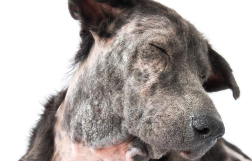
The fancy way of saying hair loss, alopecia affects more pets than we may think. It may be congenital or acquired and for it to be managed effectively, we really need to understand it. So, let’s take a look at 5 reasons for alopecia in pets. What is Alopecia? Alopecia is the partial or complete lack of hairs in areas where they are normally present. We can imagine our pet’s hair (and skin for that matter) as a report card for the body. If it’s looking a little worrisome, we need to investigate. As we mentioned, alopecia can be both congenital and acquired. Congenital means the animal is born with the condition. Congenital hair loss may or may not be hereditary. It’s caused by a lack of normal development of hair follicles. It may be apparent at, or shortly after birth. Your pet may be born with a normal coat, and patchy or widespread hair loss occurs when they become a young adult. In acquired hair loss, your pet is born with a normal hair coat. It has or had normal hair follicles at one time and is or could produce structurally normal hairs. Subsequently, any disease that can affect hair follicles can cause hair loss. Certain diseases may destroy the hair follicle or shaft or interfere with the growth of hair. Some diseases can cause discomfort, leading to self-trauma (scratching and biting) and loss of hair. It’s important to remember that acquired hair loss can be inflammatory or non-inflammatory. What diseases can Interfere with the growth of hair? Diseases that can directly cause destruction or damage to the hair shaft or follicle include bacterial, fungal, or parasitic infections. It can also include inflammatory diseases of the skin along with skin trauma. These diseases tend to be inflammatory. Parasites and What You Really Need to Know But there can also be factors that inhibit or slow down follicle growth resulting in alopecia. Let’s take a look. Nutritional Deficiencies We are seeing more and more data around specific nutrients in hair growth and health. For example: – Low vitamin D status has been implicated in cases of alopecia, – Over-supplementation of Vitamin A is associated with alopecia – In a Biotin deficiency, signs include hair loss – Folate deficiency can result in hair, skin and nail changes – Vitamin C is known to aid iron absorption, the latter being implicated in hair loss – Hair loss is a common sign of zinc deficiency – Hair loss can be seen in Iodine deficiency as it’s a mineral that aids thyroid function (we’ll share why this is relevant next) We advocate a fresh food diet, rich in nutrients to support overall health. Check out our range of blogs on different nutrients if you would like to learn more. Hormonal imbalances So much of a factor, there is a condition deemed hormonal alopecia in dogs. This can be linked to neutering with many owners reporting hair loss or thinning post neutering. But when we say hormones, we are also considering thyroid hormones. The thyroid gland is active in the initiation of hair growth and replacement. Located in the neck near the trachea or windpipe, this gland produces hormones which regulate metabolism. Both hyperthyroidism and hypothyroidism can result in hair loss in the dog although hypothyroidism is likely the more commonly occurring form of hormonal alopecia in dogs. Initially hair loss is patchy, the coat is dry, the hair is brittle and easily pulled out. Quite often hyper pigmentation occurs. In some cases, secondary pyoderma and seborrheic dermatitis may follow. Ultimate Guide: Hypothyroidism Stress Hair loss can follow months after a traumatic event often making it difficult to connect the dots. Hair cycles through different phases and all follicles can be at different stages at any one time. What we now know is that high levels of stress can cause shifts in those cycles. This results in balding or thinning of hair. Stress also depletes nutritional resources along with impeding the digestion and absorption of them and as we’ve already mentioned, sufficient growth relies on a great supply of nutrients. Can Stress Affect My Dog’s Digestive System? Irritation When your dog scratches or bites because they are irritated, it can result in hair loss. Causes of irritation include: – Infection – Pain – Parasites – Sensitivities/allergies Itchy Dogs and Cats Naturally Things to Think About: Skin Health in Dogs Overgrooming Overgrooming can be a calming behaviour employed by your pet. This may be in response to stress or being overwhelmed. Its important to notice any change in grooming behaviour and establish the potential trigger. Stressors may include: – Change in routine – Addition of a new pet – Our own stress – Change in health – Time of year – holidays/fireworks/weather change If you think hair loss may be associated with stress, check out the following blogs for more information: The Pet Owner’s Stress Load Using Nutrition To Support The Stressed Dog Why Dogs Need To Chew 5 Nutrients to Support Your Anxious Dog Overgrooming can also be linked to pain and digestive issues, so check out our blog on licking behaviour for more information. Why Does My Dog Keep Licking Signs of Hair Loss Signs of hair loss may be obvious or subtle, depending on what’s causing it. Congenital or hereditary hair loss can be symmetric (appearing similar on both sides of the body) or located in one area only. It is not usually accompanied by inflammation. Signs of acquired hair loss are influenced by the underlying causes. Hair loss may affect an isolated spot or multiple areas; it may be symmetric or widespread. You may also notice inflammation, thickened skin, colour change, scaling, excessive shedding and/or itching. In addition, some causes may lead to the development of secondary skin diseases like infection or fluid discharge. Some questions to ask when establishing the cause of your pet’s alopecia? Are they getting the nutrients they need from the diet they are offered? Could stress